
Once pure samples of DNA have been prepared, it can be manipulated in several ways; cut at specific sites and then joined together in a controlled manner. They also can be shortened, lengthened, copied into RNA or into new DNA molecules and modified by the addition or removal of specific chemical groups. These manipulations, all of which can be carried out in the test tube, provide the foundation not only for gene cloning, but also for basic studies into DNA biochemistry, gene structure, and the control of gene expression.
The cutting and joining manipulations that underlies gene cloning are carried out by enzymes called restriction endonucleases (for cutting) and ligases (for joining).
Nucleases degrade DNA molecules by breaking the phosphodiester bonds that link one nucleotide to the next in a DNA strand. There are two distinct kinds of nucleases:
During this week you will carry out restriction digestion of the genomic clone, in order to cut the insert (=the gene) from the plasmid DNA (=vector). Following separation of insert and vector by electrophoresis, you will elute the insert from the agarose. More background information about agarose gel electrophoresis and related techniques (e.g., blotting) can be found at these links. Detection of DNA on the gel can be manifested by the presence of Ethydium Bromide in the gel which binds or intercalate and the resulting molecule fluoresce when illuminated with UV light. Gel electrophoresis is frequently performed with marker DNA fragments of known size which allow accurate size determination of an unknown DNA molecule by interpolation.
After performing the electrophoresis, the piece/band in the gel corresponding to the isolated FABP insert will be cut off the gel and the DNA fragment will be eluted from the gel. The eluted DNA will then be used for DNA amplification by the Polymerase Chain Reaction (week 3)
1. Prepare the restriction enzyme digests. You will perform two single digests using enzymes EcoRI and HindIII and one double digest using both enzymes. You will also be given some plasmid prepared by your TA to use as a positive control. Prepare one single digest (choose either EcoRI or HindIII) and one double digest for this plasmid as well.
For each of the single digests you will add:
( ) µl sterile ddH20
2 µl 10X reaction buffer - REact 3 for EcoRI or REact 2 for HindIII
( ) µ plasmid DNA - calculate ~20-25 µg of DNA, but don't add more than 10 µl
3 µl of 10 U/µl restriction enzyme - 1 Unit cleaves 1 µg of DNA in one hour at 37 °C
__________________________
25 µl total reaction volume
For each of the double digests you will add:
( ) µl sterile ddH20
2 µl 10X reaction buffer - choose either REact 3 or REact 2
( ) µl plasmid DNA - calculate ~20-25 µg of DNA, but don't add more than 10 µl
3 µl of 10 U/µl EcoRI restriction enzyme - 1 Unit cleaves 1 µg of _ DNA in one hour at 37 °C
3 _l of 10 U/_l HindIII restriction enzyme - 1 Unit cleaves 1 µg of _ DNA in one hour at 37 °C
__________________________
25 _l total reaction volume
2. Incubate at 37 °C for 1 hour. If you are using a heating block, make sure the wells have added water to promote even heat transfer. Proceed with setting the agarose gel in Step 3.
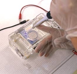
3. Set up a gel tray as demonstrated by your TA. Then, to make a 1 % agarose gel, weigh 0.5 g of agarose and add to 50 ml of 1X TAE (Tris-EDTA-Acetic acid, pH 8.0) in a clean 250 ml Erlenmeyer flask.
4. Microwave at high setting for 40 s. Swirl the solution and microwave for an additional 10 s.
5. Add 5 µl of 10000X GelRed. GelRed is an intercalating DNA stain that works in the same manner as ethidium bromide except it much safer to use and is slightly more sensitive. Swirl to mix and pour the agarose into the mold and insert an 8-well comb. It will take approximately 20-30 min to set.
6. After the incubation period, take the digested samples and add 4 µl of 6X loading buffer to each tube. Also, add 3 µl loading buffer to 10 µl aliquots of undigested plasmid, control plasmid given to you by your TA. Mix well by flicking on the side. Spin in the centrifuge at high speed for 10 s to gather the contents to the bottom.
7. Prepare the run buffer (1X TAE pH 8.0). Place the gel in the gel box. Pour the buffer to cover the wells.
8. Using a pipetman, load
the gel. Load your samples with the following amounts: 1 Kb Fermentas O'GeneRuler DNA Ladder (6 µl it is already in loading buffer), your TA's undigested R32 plasmid (13 µl), the single digest from your TA's plasmid (24 µl), the double digest from your TA's plasmid (24 µl), your undigested plasmid (13 µl), your single digests (24 µl) and your double digest (24 µl).
9. Electrophorese at 100 V for 1 hour or until the lighter portion of the dye front is 1 cm from the bottom of the gel.
11. We will be using the GE Illustra GFX PCR and Gel Band Purification kit to elute the DNA from the gel slice containing your R32 insert. While the gel is running, pre-weigh a sterile Eppendorf tube in which you will collect the gel slice containing the desired DNA fragment. Also, obtain and label a spin column and collection tube for the gel extraction procedure. Make sure a heat block is set to 60 °C.
12. Once the electrophoresis is finished, turn off the power pack and disassemble the gel set up. Observe under UV light using the gel documentation system in B7179. Take a picture of your gel - be sure to both print and save a copy.
13. Cut the appropriate band from your gel using the table-top transilluminator (wear a UV-safe mask). Cut the gel as close to the DNA band as possible. Transfer the gel-slice to the pre-weighed Eppendorf tube.
14. Re-weight the Eppendorf tube and determine the weight of the gel slice. For every 1 mg of gel, add 1 µl of GFX Capture Buffer. Do not add more than 300 µl.
15. Incubate the tube on the heat block for 5-15 min until the gel is melted. Be sure to vortex the tube several times during the incubation.
16. Once the agarose is completely dissolved, transfer the solution to the GFX spin column and let sit for 1 minute at room temperature.
17. Spin the column at maximum speed for 1 minute.
18. Discard the flow-through by emptying the collection tube (do not throw the tube away!). Add 500 µl Wash Buffer (with ethanol already added).
19. Spin the column at maximum speed for 1 minute. Discard the flow-through and spin again for another 30 s to ensure there is no residual buffer in the spin column.
20. Transfer the spin column to a sterile 1.5 ml Eppendorf tube. Add 50 µl of sterile 1X TE buffer or sterile ddH20 to the centre of the column. Let sit 1 minute.
21. Spin the column at maximum speed for 1 minute and then discard the column. The flow-through contains your eluted DNA.
22. If there is time, you will receive a demonstration of how to use the NanoDrop spectrophotometer as a good measure to determine the concentration and purity of your eluted DNA sample (the NanoDrop is an alternative to the standard spectrophotometer you used to measure your DNA in the previous week's lab). Use the same buffer/water you used to elute your DNA from the spin column as a blank.
![]()
![]()
[
BISC 429 home]
[
Enzyme isolation ] [
Lipoprotein isolation ] [
Lipid analysis ] [
DNA
isolation ] [ feedback ]