
Overview of the circulatory system of the shark. |
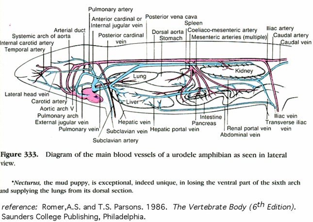
Overview of the Circulatory System of a Salamander. |

Overview of the Circulatory System of the Cat. |

Diagram of blood circuits and capillary networks in a typical A. fish, B. amphibian or reptile and C. a mammal. |

Diagram of lateral and dorsal views of the evolution of posterior cardinals and the development of the vena cava. |

Diagram showing the evolution of the Aortic Arches in Representative Vertebrate Groups. |

The Evolution of the Aortic Arches in Vertebrates. |

Diagram showing the Evolution of Major Venous Systems in Representative Vertebrate Groups. |

Diagram showing the Evolution of Venous Return Circuits in Shark, Amphibian and Mammal. |

|

Shark Arteries in the Abdominal Cavity, Ventral View, Labelled. |

|
 Shark Arteries Associated with the Gills and the Snout, Ventral View, Unlabelled. |
 Shark Arteries Associated with the Gills and the Snout, Ventral View, Labelled. |

Shark Heart ,Ventral View - Unlabelled. |

Shark Cranial Arteries. Ventral view of the dorsal aorta with the skin on the roof of the mouth intact. |

Shark Cranial Arteries. Ventral view of the ventral aorta and afferent branchial arteries. Note: These blood vessels are latexed blue, whereas in our specimens, these blood vessels will not be latexed. |
 Shark Heart ,Ventral View - Labelled. |

Diagram of the Major Arteries and Veins Associated with the Heart and Gills in Shark. |

Black and White Diagram of the Heart of the Shark, Longitudinal Section. |

Shark Heart, Ventral View with Ventricle Pulled to Side to Expose Sinus Venosus, Labelled. |

Black and white diagram of the structure of the gills of the shark. |

Diagram of the flow of blood through the gill of a shark. Red is oxygenated blood. Blue is deoxygenated blood. |

Shark Heart, Ventral Veiw with Ventricle |

Ventral View of the head of the shark, showing the inferior jugular vein and heart, Unlabelled. |

Ventral view of the head of the shark, showing the inferior jugular vein and heart, Labelled. |

Ventral view of the blood vessels associated with the kidney, Unlabelled. |
 Ventral view of thoraic cavity of shark, Unlabelled. |
 |

Ventral view of the blood vessels associated with the kidney, Labelled. |

Ventral and Dorsal Views of the Heart of the Turtle. |
 |
 |

Ventral View of a Mammal Heart. |

Sectional Diagrams of Amphibian Heart. |

Colour Diagram of the Heart of Necturus. |

Ventral view of Necturusheart and ventral aorta, Unlabelled. |

Ventral view of Necturus heart and ventral aorta, Labelled. |

Ventral view of Necturus Circulatory System, Unlabelled. |

Ventral view of Necturus radix aorta and dorsal aorta, Unlabelled. |

Ventral view of Necturus radix aorta and dorsal aorta, Labelled. |

Ventral view of Necturus Circulatory System, Labelled. |

Ventral view of the heart and associated blood vessels of Necturus, Unlabelled. |

Ventral view of the heart and associated blood vessels of Necturus, Labelled. |

Ventral view of the heart and blood vessels anterior to the heart of Necturus, Labelled. |

Vental view of the heart and associated blood vessels of Necturus, Labelled. |

Ventral view of the radix aorta of Necturus, Unlabelled. |

Ventral view of the radix aorta of Necturus, Labelled. |

Ventral and Dorsal Views of the Origin of Major Blood Vessels Associated with the Heart of the Turtle. |
 |

Vental View of the Renal Portal System of the Turtle. |

Ventral View of the Arteries of the Turtle. |

Ventral View of the Veins of the Turtle. |

Selected arteries of a Pigeon. |

Ventral view of the arteries of the turtle. Reference: Hildebrand, M. and G. Goslow 2001, Analysis of Vertebrate Structure 5th edition. John Wiley and Sons, Inc., New York |

Ventral view of the veins of the turtle. Reference: Hildebrand, M. and G. Goslow 2001, Analysis of Vertebrate Structure 5th edition. John Wiley and Sons, Inc., New York |

Selected veins of a Pigeon. |

Abdominal blood vessels of pigeon, Unlabelled. |

Abdominal blood vessels of pigeon, Labelled. |

Ventral view of the male pigeon urogenital system, Unlabelled. |
 Ventral view of the renal portal vein of the pigeon, Unlabelled. |

Ventral view of the renal portal vein of the pigeon, Labelled. |

Ventral view of the male pigeon urogenital system, Labelled. |

Ventral view of the heart and associated blood vessels of a pigeon, Unlabelled. |

Ventral view of the heart and associated blood vessels of a pigeon, Labelled. |

Ventral View of an Avian Heart. |
 Ventral View of the turtle heart and associated blood vessels. Liver included in the photograph. Unlabelled. |
 Ventral View of the turtle heart and associated blood vessels. Liver included in the photograph. Labelled. |
 Turtle heart showing sinus venosus and the 4 veins which bring deoxygenated blood to it. Unlabelled. |
Turtle heart sinus venosus with hepatic vein and the posterior vena cava. Unlabelled. |
 Turtle heart sinus venosus with hepatic vein and the posterior vena cava. Labelled. |
 Turtle heart showing sinus venosus and the 4 veins which bring deoxygenated blood to it. Labelled. |
 Turtle heart sinus venosus and left anterior vena cava. Left pulmonary vein also visible. Unlabelled. |
 Turtle heart sinus venosus and
left anterior vena cava. Left pulmonary vein also visible.
Labelled. |