Medical research project to capture 3D images during surgery
Dr. Ghassan Hamarneh from SFU Computing Science is leading a research project to benefit surgeons and their patients. His research team aims to bring the rich details of pre-operative medical images to surgeons who perform robotic surgeries by capturing and utilizing dynamic, 3D images indicating changes in a patient’s body during the course of an operation.
Dr. Hamarneh is the lead investigator on a project that received $1 million US from the Qatar National Research Foundation. He is working closely with the University of British Columbia, The Cleveland Clinic and the Qatar Robotic Surgery Centre to acquire, analyze, and visualize 3-D anatomical images in real-time. Dr. Hamarneh foresees this funding as the first step towards building a strategic alliance with Qatar’s emerging research and technology sector in medical image computing.
Robotic surgery is the latest in what is known as minimally invasive surgeries involving smaller incisions and it offers many advantages such as reduced blood loss, and a lower chance of infection and complication. During robotic surgery, the surgeon sits down away from the patient at a special console where he accesses patient images and where he controls precise movements using remote-controlled “arms.” Today, the surgeon is located in the same room as the patient but in the future, being in a different location may become increasingly common.
“Robotic surgery is the key technology for tele-surgery and future unmanned surgeries, so it is poised to become one of the most important forms of surgery in the coming decades,” said Dr. Hamarneh, an associate professor with SFU Computing Science.
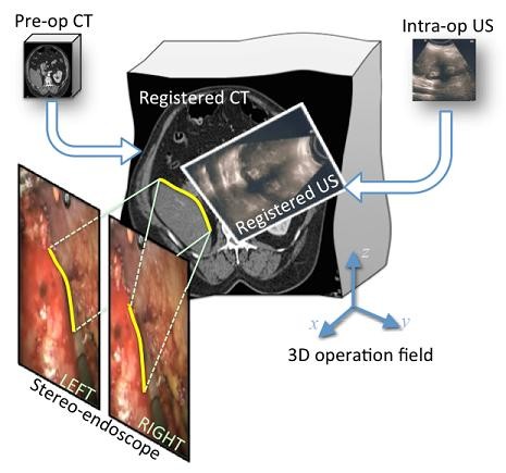
According to Dr. Hamarneh, some of the challenges involved include ensuring images taken during surgery can be processed very quickly, automatically and accurately, and are able to cope with a highly dynamic surgical scene. Relevant anatomical structures in the images must be identified automatically (a process called image segmentation) and images from different sources (e.g. a pre-operative CT scan and an ultrasound taken during surgery) must be brought into proper alignment (image registration). The project will focus on developing such techniques through advanced computer vision, 3D image and geometry processing methods, as well as optimization and machine learning algorithms.
Modern medical imaging technologies are revolutionizing medicine, and Dr. Hamarneh and his team of researchers will be combining the best imaging technology to advance real-time patient information.
For more information, contact fascomms@sfu.ca.
Comment Guidelines