New laser scanners shed light on eye disease before vision loss occurs
By Diane Luckow, SFU News
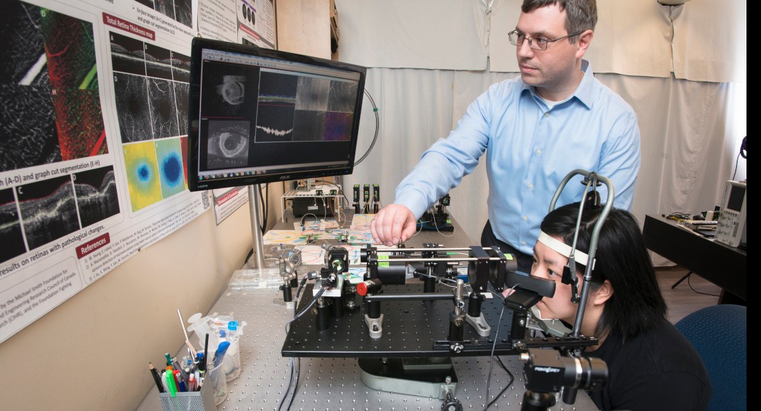
SFU engineering scientist Marinko Sarunic has developed new retinal imaging scanners that could revolutionize eye care, helping ophthalmologists diagnose eye diseases before vision loss occurs.
The retina is the light-sensitive tissue at the rear of the eye. Its 100 million photoreceptors convert light into the images that our brain ‘sees.’
Sarunic’s high-resolution laser scanner is on the cutting edge of vision science because it can produce high-resolution, 3-D cross-section images of the retina—including individual photoreceptors, and fine capillaries, or blood vessels.
And unlike other high-resolution retinal scanners, which are the size of a billiard table, Sarunic’s is the size of a shoe box. It’s perfect for everyday use in medical clinics and hospitals.
“It’s a breakthrough in clinical diagnostics,” says Sarunic. “With the high-resolution scanner, ophthalmologists and optometrists can detect damage and changes to small numbers of individual photoreceptors, giving them a diagnosis before the patient loses vision, and the potential to take preventative measures.”
And since the technology is not invasive, he says physicians can use the scanner frequently to monitor the retina for changes or to detect whether medications are working.
Currently, physicians must use low-resolution scanners, which can only assess and diagnose the cause of dead retina cells after a patient has lost vision.
Last year, ophthalmologists at Vancouver General Hospital’s (VGH) Eye Care Centre spent eight months testing Sarunic’s high-resolution scanner.
Dr. Eduardo Navajas, a vitreoretinal specialist, says the scanner eliminates the need for, and the complications related to, dye injections that are currently used to diagnose and monitor eye diseases like diabetic retinopathy and wet age-related macular degeneration (Wet AMD). These are the two most common causes of vision loss.
“Early detection of abnormal blood vessels caused by Wet AMD and diabetes is essential to saving a patient’s vision,” says Navajas. “Dr. Sarunic’s new imaging technology is benefiting patients, allowing us to diagnose and treat Wet AMD and diabetic eye disease before patients develop bleeding and permanent damage to their retina.”
Sarunic is now developing another version of the scanner that ophthalmologists can use for image-guided operations.
“As data is pulled off the scanner it presents the results to the physician, providing guidance during surgery,” he says. “As they operate on eye tissue or do laser-based changes, they can track what they’re doing.”
Sarunic’s research stems from previous jobs in industry, where he developed fibre optics projects for telecommunications companies. He joined SFU in 2006 to pursue applied research that could have real impact on peoples’ health.
“I wanted to do something more meaningful,” he says. “I find it very rewarding to work with people to address their needs.”
He is now working with the SFU Innovation Office to commercialize his scanner technology.
He conducted his research with grants from: Michael Smith Foundation for Health Research (MSFHR), National Science and Engineering Research Council (NSERC), Canadian Institutes of Health Research (CIHR), Alzheimer Society of Canada, Pacific Alzheimer Research Foundation, Brain Canada, Genome Canada, and the Foundation Fighting Blindness.