ApoC-II
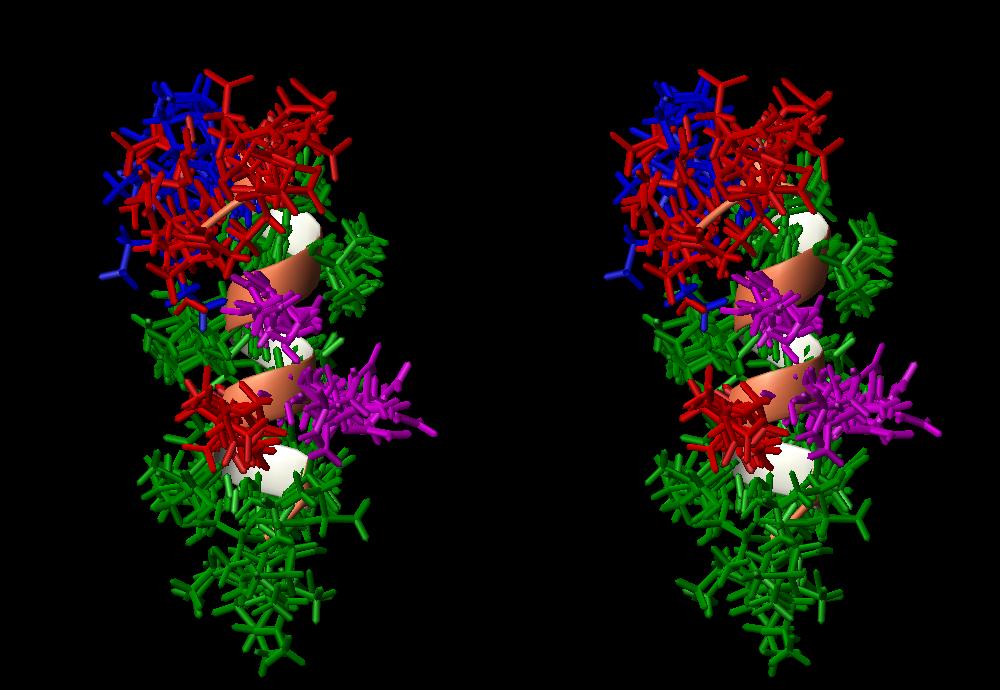
Caption: Preliminary stereoview of the primary lipid binding domain of apoC-II(44-79) which is composed of residues 66 through 75 as determined from NMR. Hydrophobic residues are green, acidic residues are red, basic blue and hydroxyl residues (Ser and Thr) purple. The amphipathic character of the helix is clearly visible, with the hydrophillic face towards the front. The biologically important region 76-79, sequence Lys-Gly-Glu-Glu forms a turn which can be seen at the top of the figure.
Created by Kathy Cushley
last modified: June 1, 1998
URL: http://www.sfu.ca/~cushley