APOLIPOPROTEIN C-I

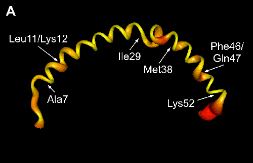
Ribbon representation of the structure closest to the mean coordinates of 18 calculated structures of apoC-I in a lipid-mimetic environment. The width and color of the ribbon is modified to reflect the circular variance of the torsion angle phi. Red shading and greater widths indicate poorly defined regions. The amino acid labels mark the locations of the N- and the C-terminal helices and the positions of highest circular variance within them, which are also the sites of bends.
Brookhaven Protein Data Bank accession number is 1IOJ.
Dr. Robert J. Cushley
Biological NMR Group
Simon Fraser University
Institute of Molecular Biology and Biochemistry
Burnaby, British Columbia, Canada V5A 1S6
cushley@sfu.ca
Institute of Molecular Biology and Biochemistry,
Simon Fraser University
Created by KathyCushley
last modified: February 28, 2000
URL: http://www.sfu.ca/~cushley

