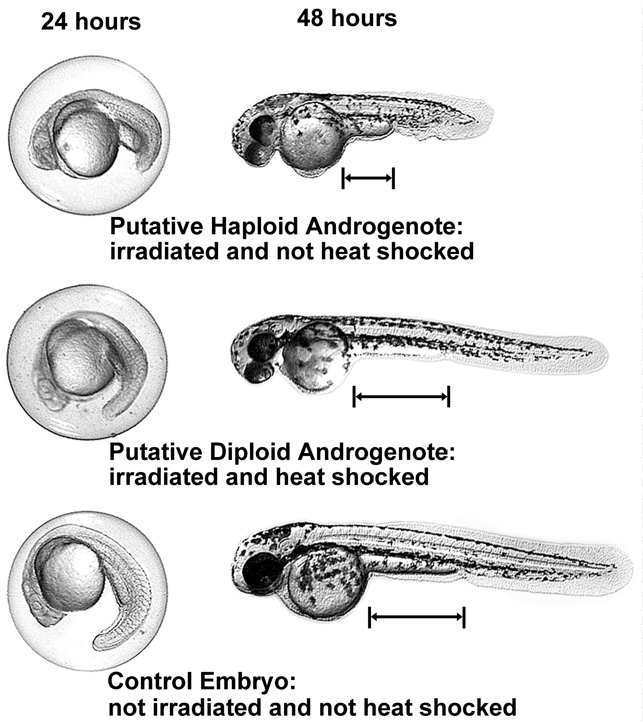
Figure 1: Comparison of haploid and diploid androgenotes and normal
diploid embryos at 24 and 48 hours. Three embryos are shown, each
at two different stages of development. Note: the distance between
the posterior yolk sac margin and the anal pore is greater for
the diploid phenotype than for the haploid phenotype.
Return to Androgenesis Protocol