Research focus
- Computational Brain Anatomy
- Computational Eye Anatomy
- Image Registration and Segmentation
- Image Acquisition
- Quality Check Visualizations
- Sponsors & Funding
- Manually segmented template library for 8-year-old pediatric brain MRI data with 16 subcortical structures
- Pediatric Shape Analysis (under review)
- Shape Analysis pipeline (in preparation)

Computational Brain Anatomy
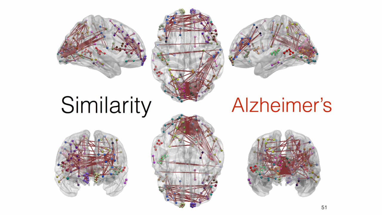
Network Analysis

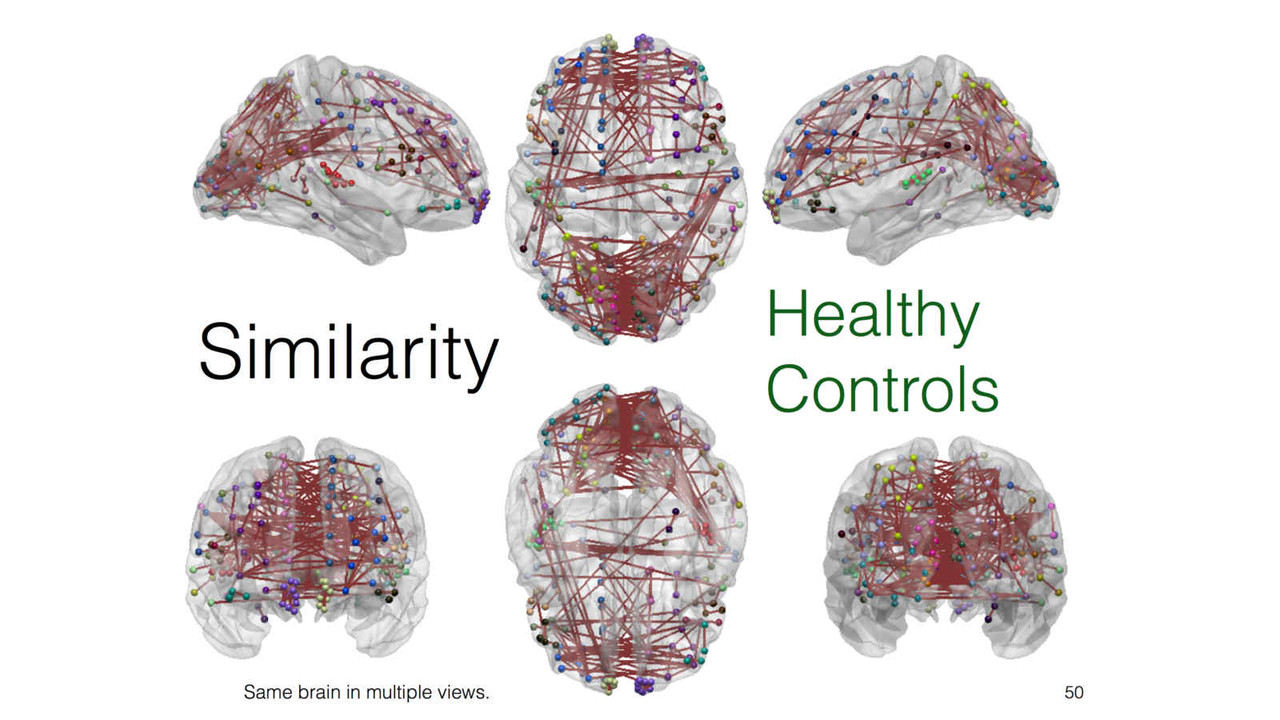
In this stream of research, we explore and investigate novel methods of extracting new insights into a given feature such as cortical thickness, via network-level analysis. Analyzing the inter-regional covariance in a given feature povides with us a richer description of the underlying patterns, and network-level changes caused by neurodegenerative diseases such as Alzheimer's disease.
Related publicationsImaging Genetics
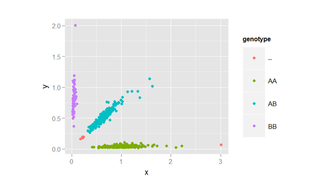
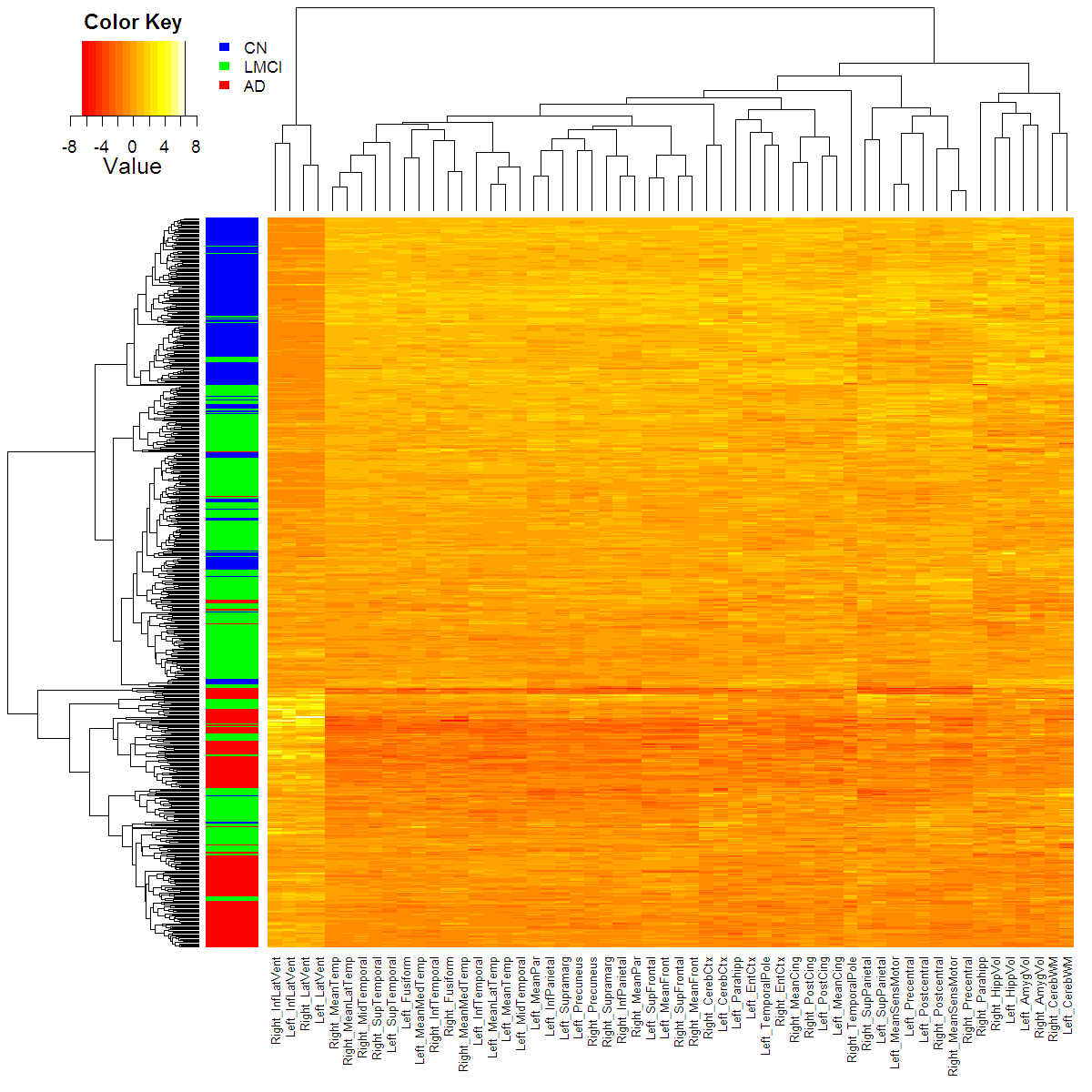
Both genetic variants and brain region abnormalities are recognized to play a role in cognitive decline. In this project, we explore the relationship between genome-wide variation and region-specific rates of decline in brain structure, as measured by magnetic resonance imaging.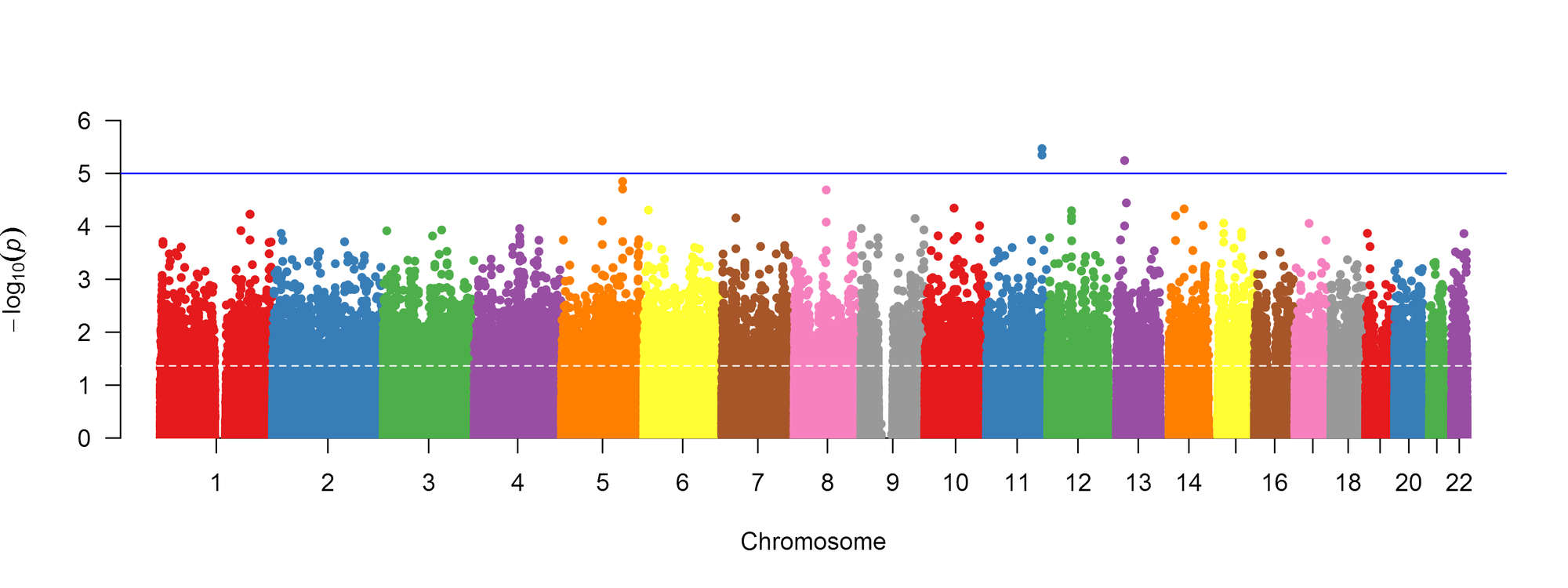
Above: Manhattan plot of association between genetic markers and cognitive impairment, adjusted for confounders.
Below: Heatmap of the rates of change in MRI features with coloured disease labels, adjusted for confounders.
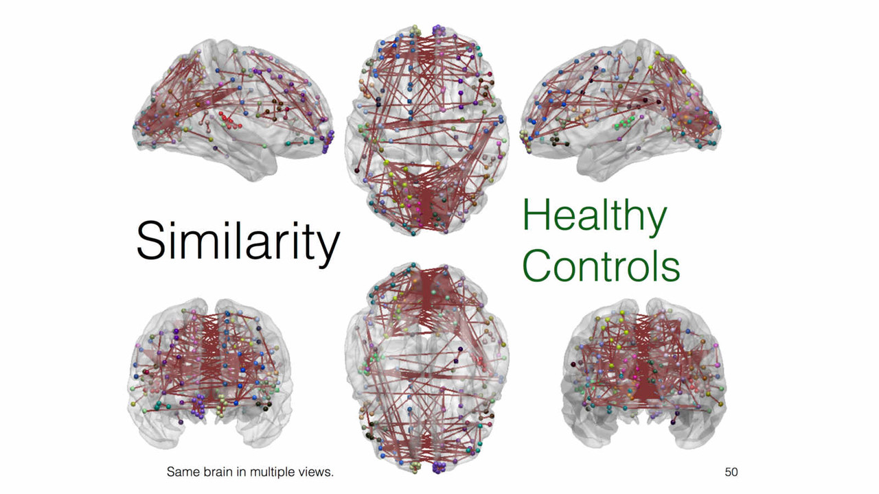
Alzeheimer's Disease
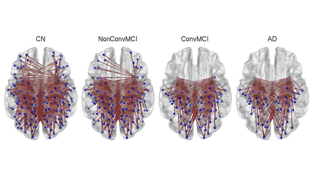
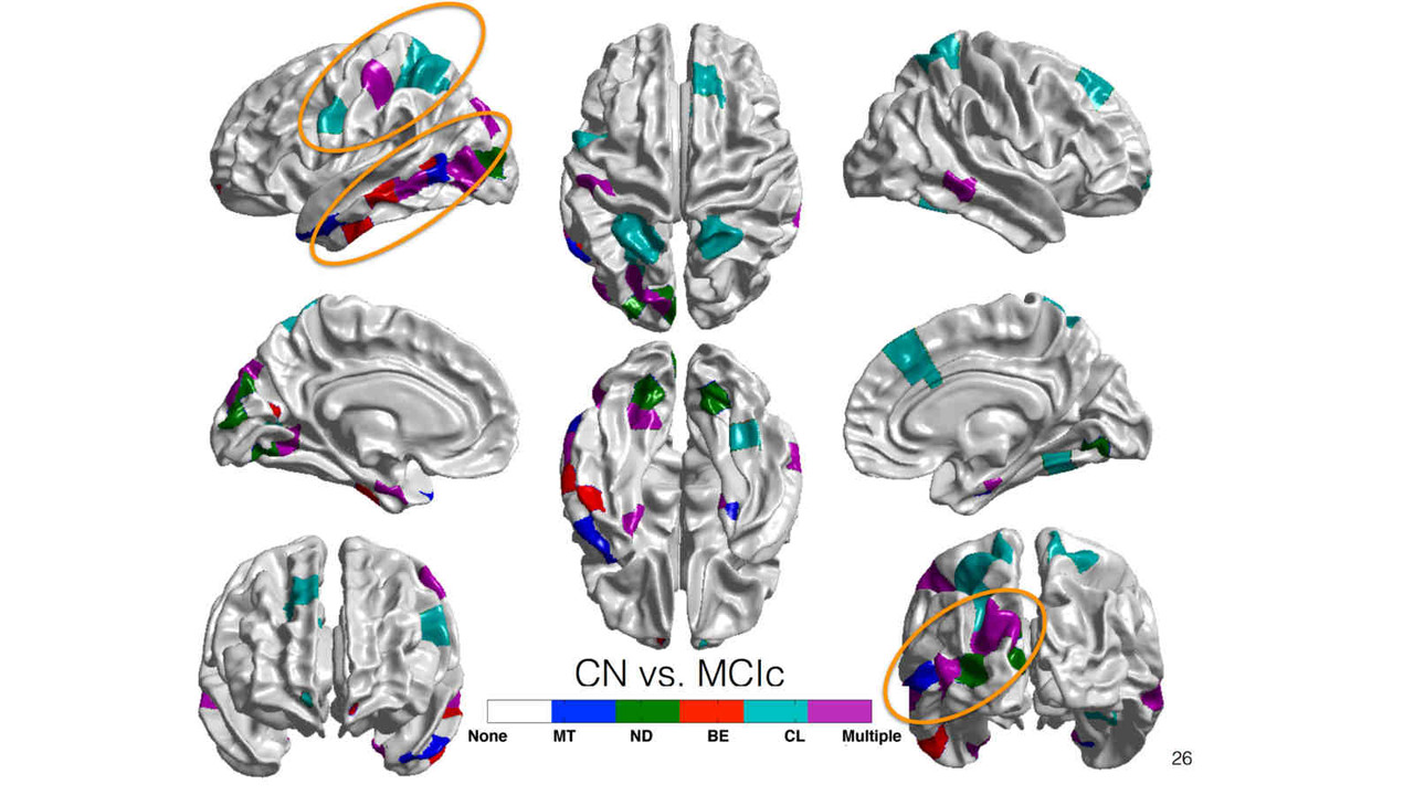
A key focus of our laboratory is on the development of fully automated computational-anatomy tools for the early detection of Alzheimer's disease. Towards this challenging goal, we develop various method through the spectrum of medical image analysis starting from organ segmentation, accurate registration, feature extraction methods and machine learning tools. The above picture shows a comparison of novel T1-MRI based networks we have constructed to help build new covariance features which show promise for the early detection of Alzheimer's disease. Please visit pages in the Research section to learn more about our research.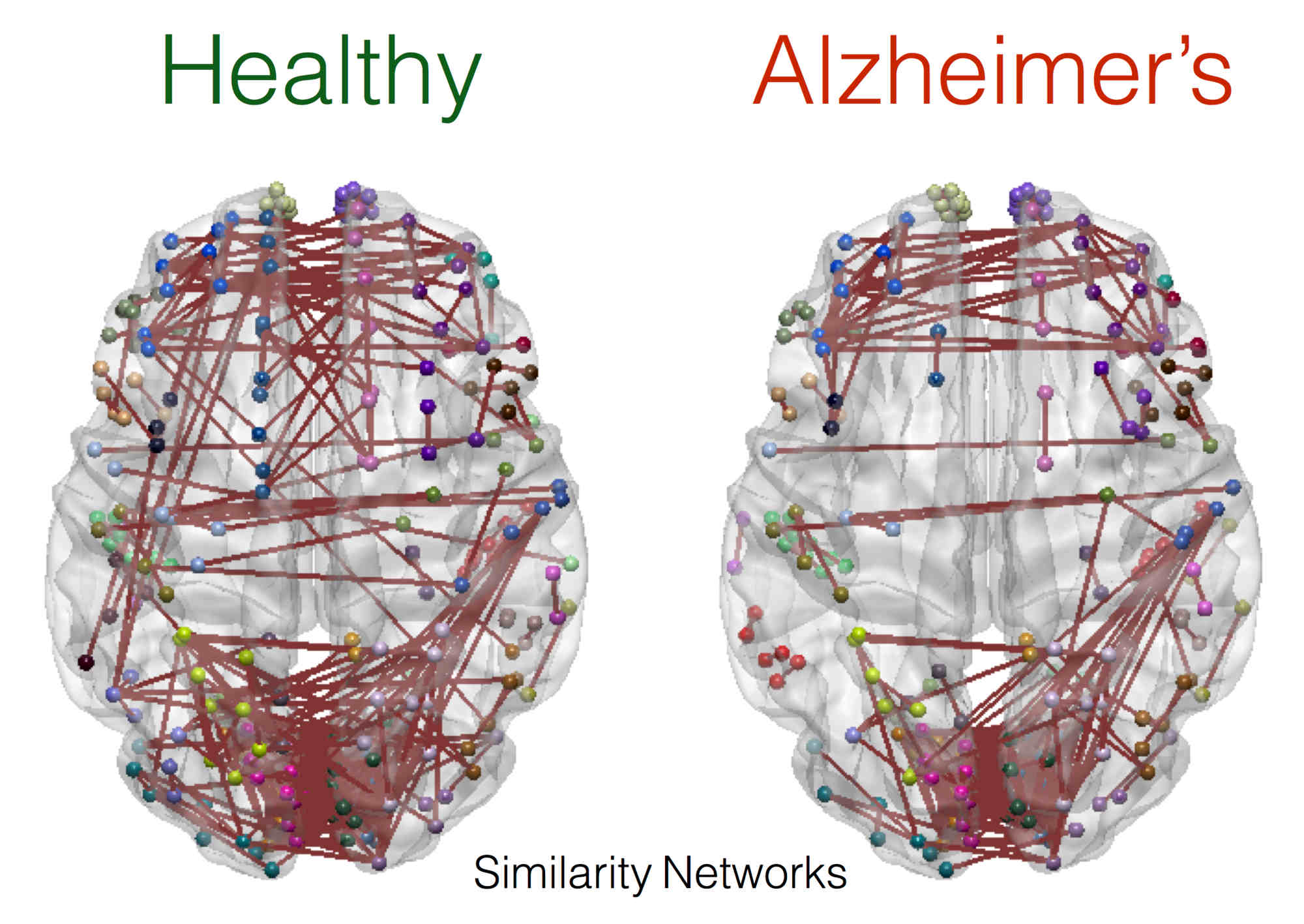
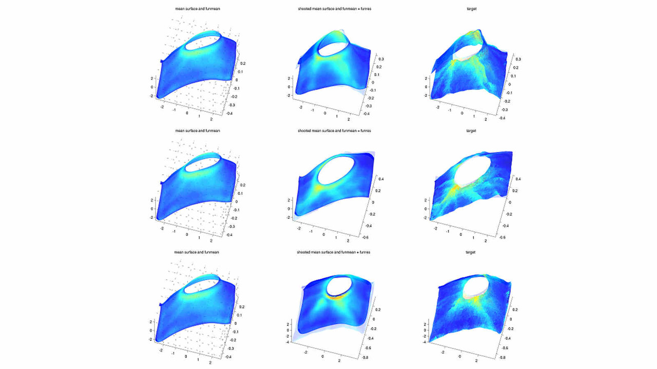
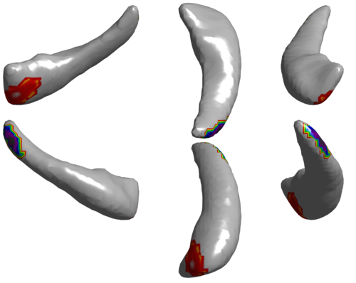
Functional Shape Analysis
 In this project, we employ the novel functional shape analysis (fshape) paradigm for the characterization of shape variability in cortical and subcortical regions extracted from MRI images in the brain, among healthy individuals, and patients suffering with Alzheimer's disease (AD) and related dementias.
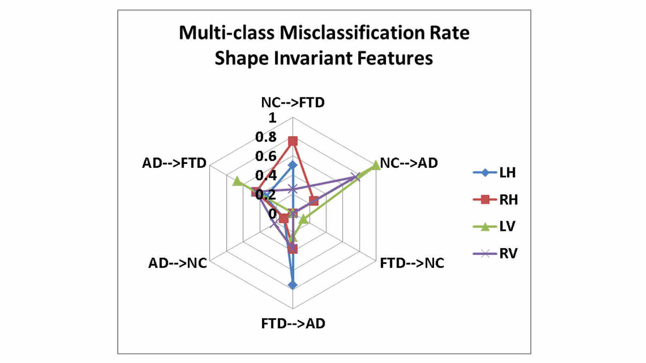
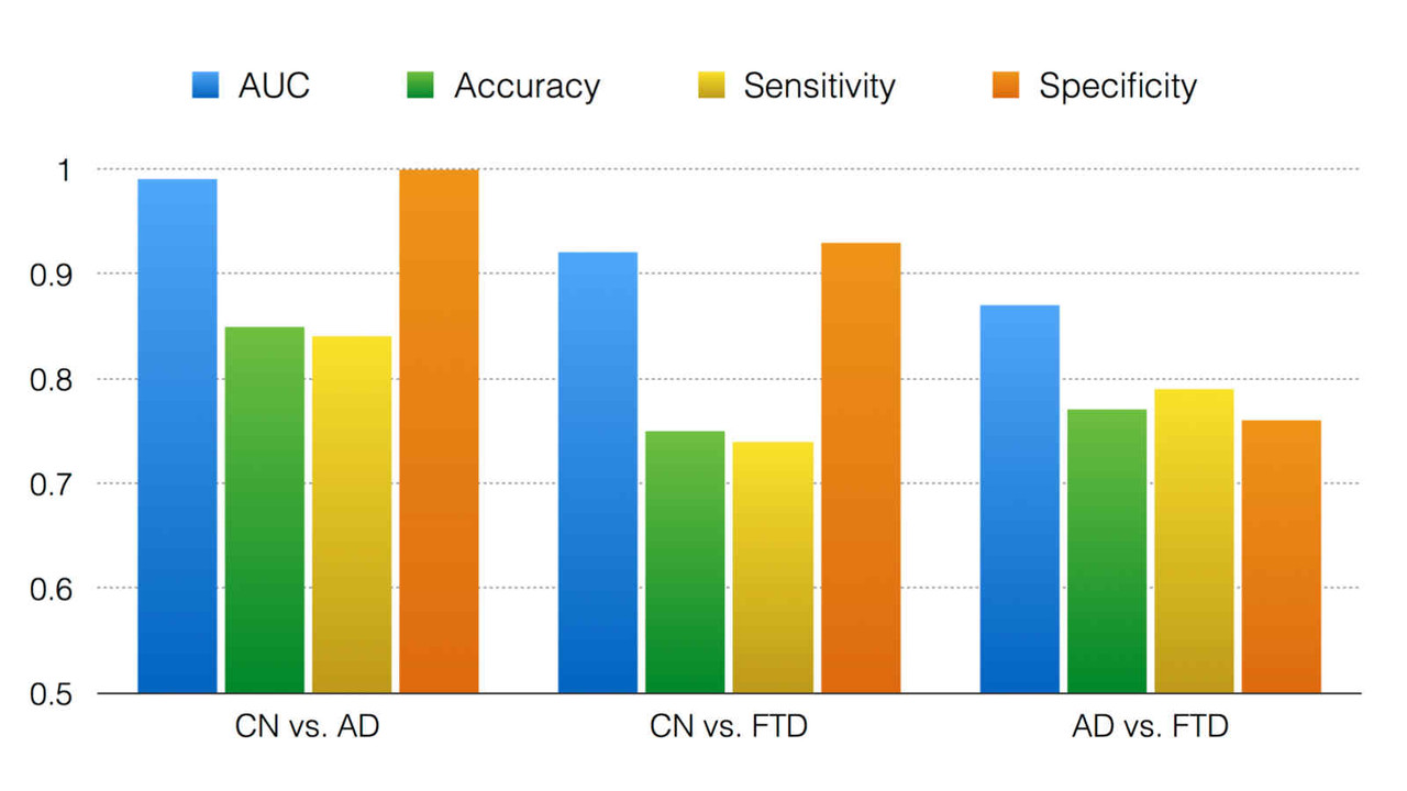
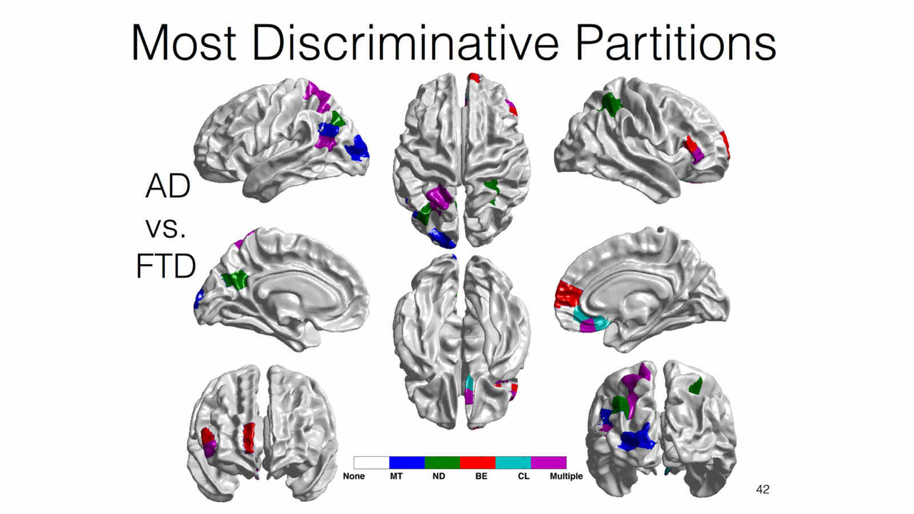
Differential Diagnosis
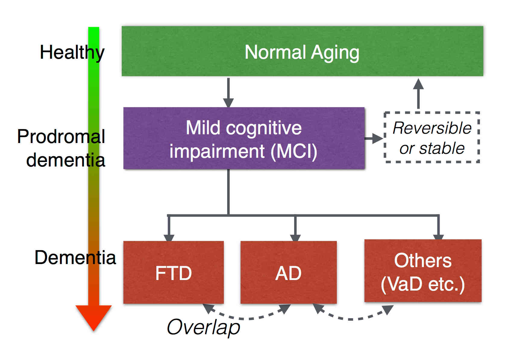
Biomarkers derived from brain magnetic resonance (MR) imaging have promise in being able to assist in the clinical diagnosis of brain pathologies. These have been used in many studies in which the goal has been to distinguish between pathologies such as Alzheimer’s disease and healthy aging. However, other dementias, in particular, frontotemporal dementia, also present overlapping pathological brain morphometry patterns. In this stream of research, we investigate novel methods for the differential diagnosis of various neurodegenrative diseases in a multi-class setting, and explore novel methods and techniques to improve the differential diagnostic accuracy.
Related Publications:Pradeep Reddy, Howard Rosen, Bruce Miller, Michael Weiner, Lei Wang, Mirza Faisal Beg, Three class differential diagnosis among AD, FTD and controls, Frontiers in Neurol- ogy, 5(71), 2014.
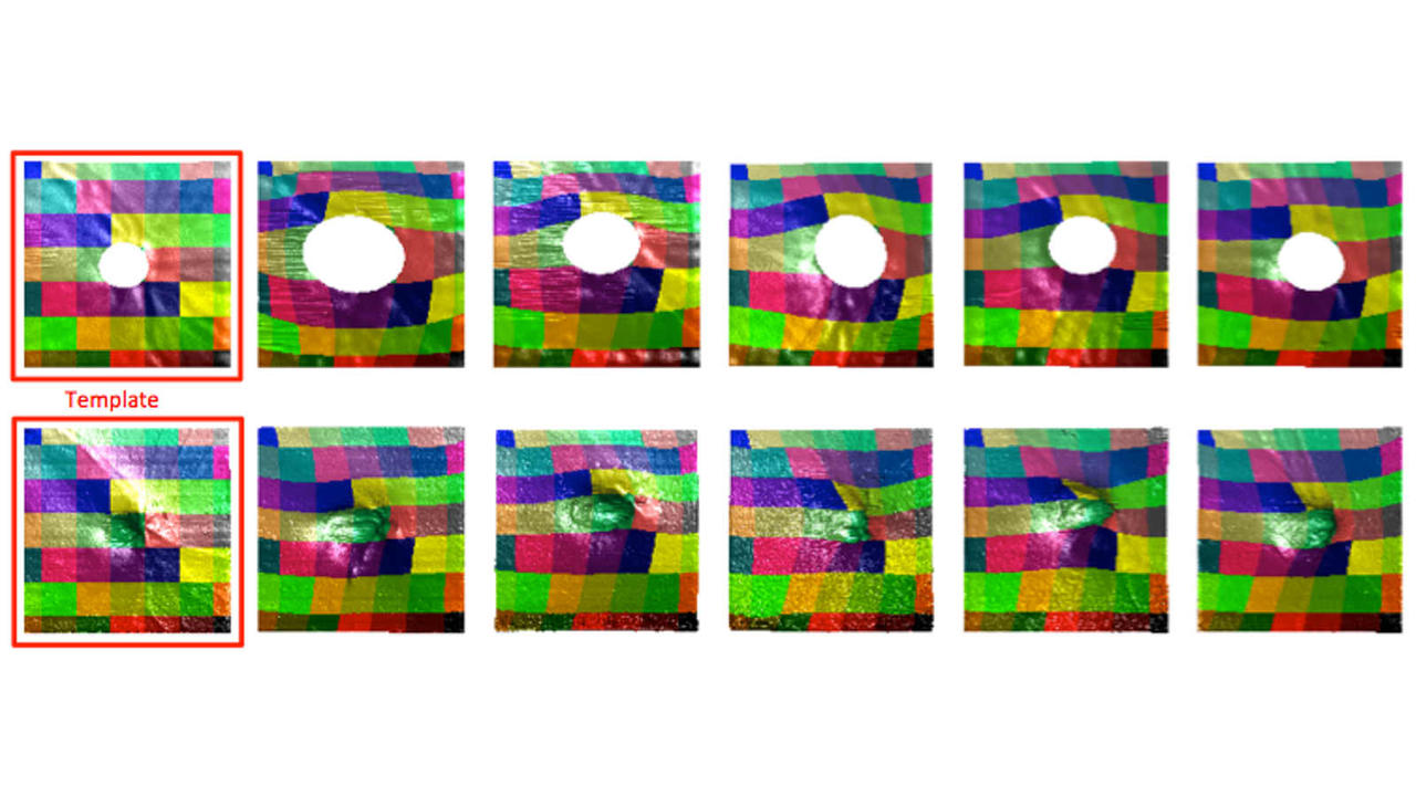
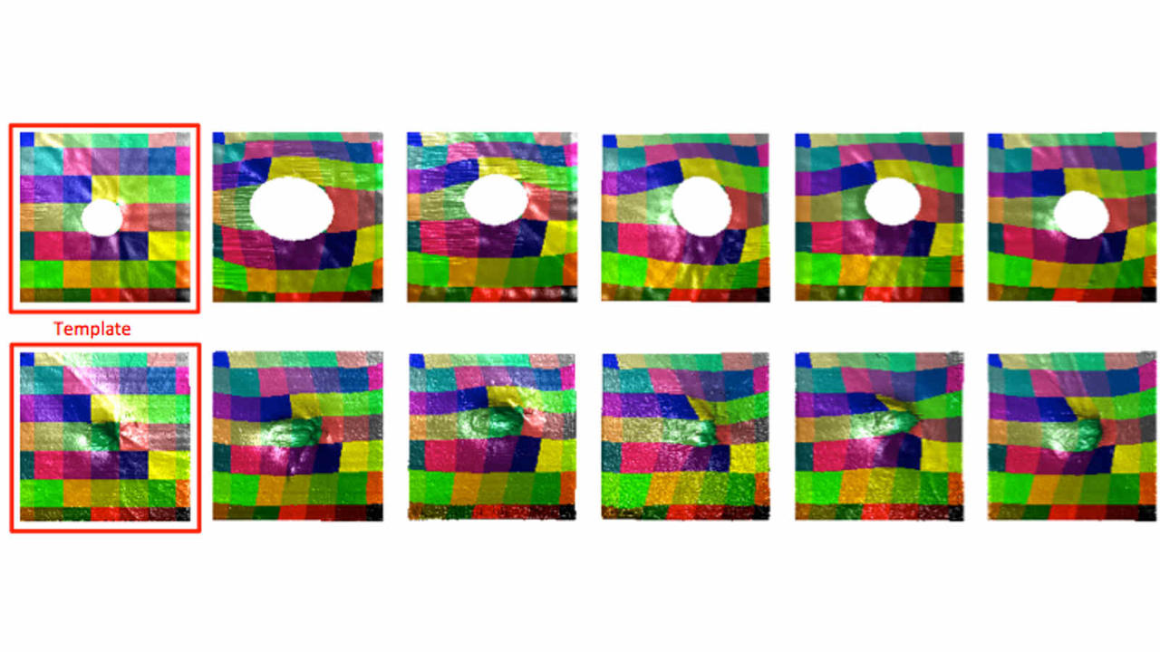
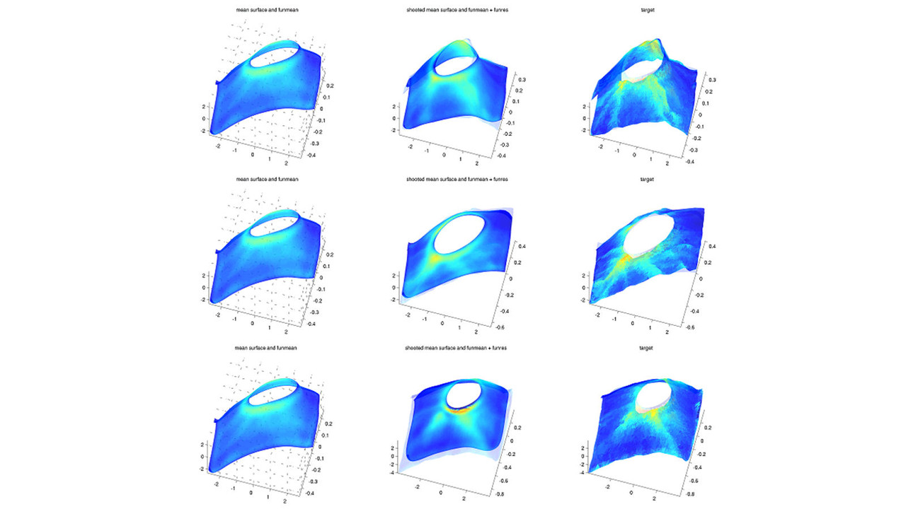
Pediatric Shape Analysis
 The change in shape of the anatomical structures within the brain is expected to have a profound effect on behavioral, learning and psychological outcomes. Brain development during early childhood from birth to adolescence is accompanied with increase in brain volume, neuronal growth in the cortex and shape formation of structures within the brain. In this work we aim to develop an automated method for analysis of brain MRI data and objective quantification of the change in shape within the brain with normal growth and the associated alterations due to diseased conditions.
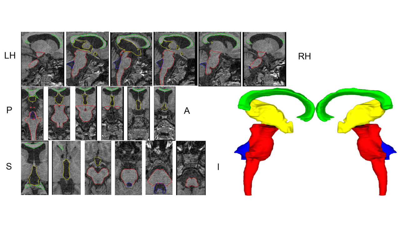
Pediatric Template Library
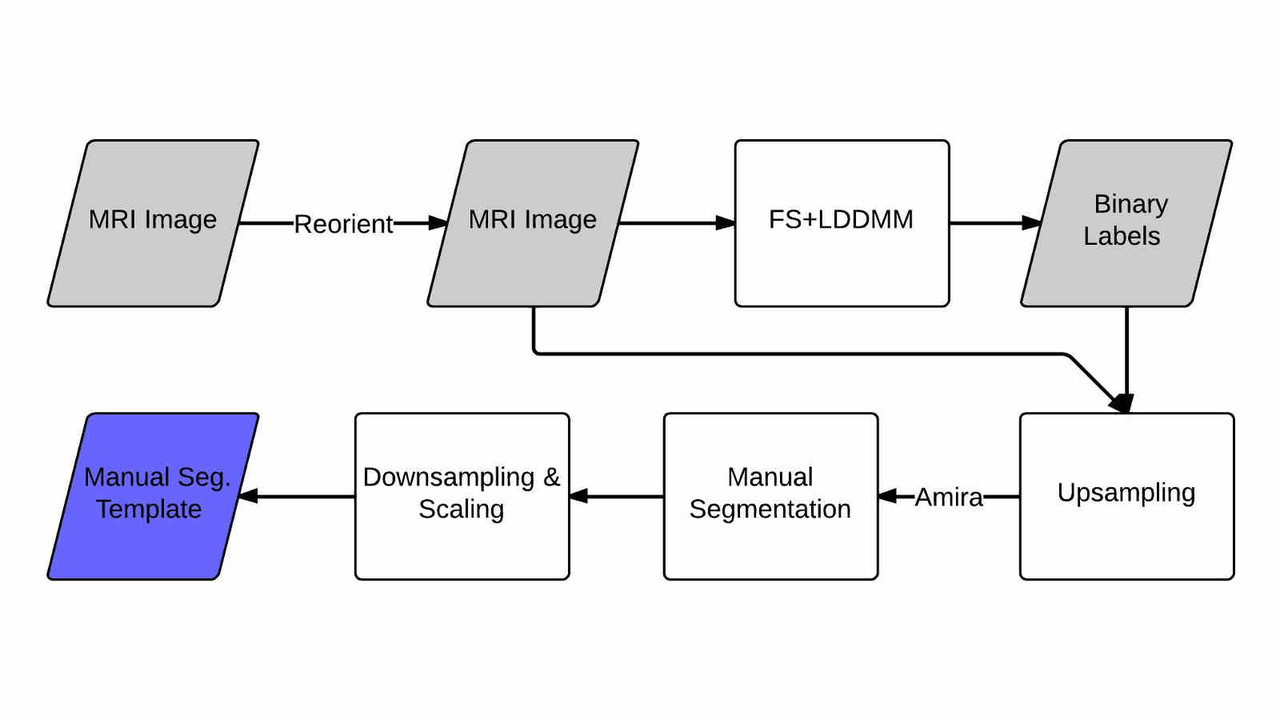
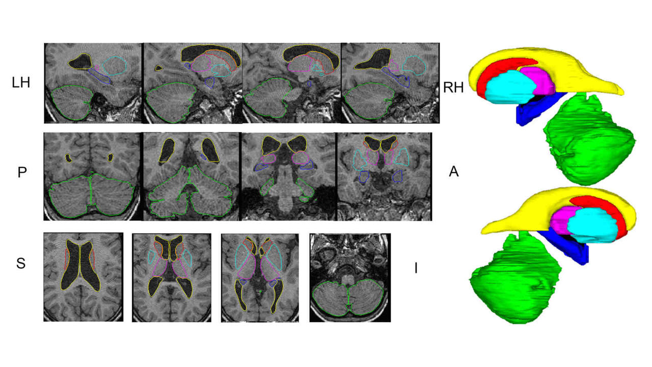
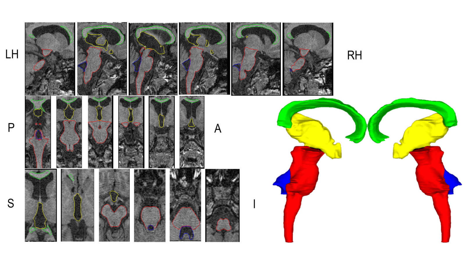
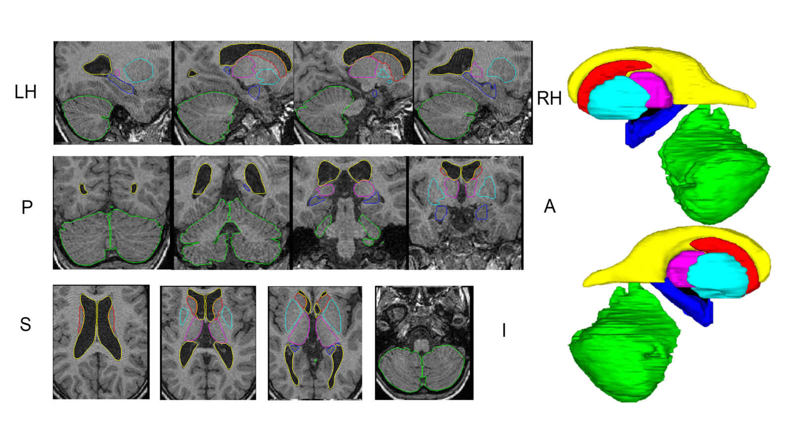
In this work we created a library of ground truth template data with manual segmentation labels for 16 subcortical structures in brain MRI data from children at 8-years of age.Publication: Manually segmented template library for 8-year-old pediatric brain MRI data with 16 subcortical structures
 Above: The manual segmentation procedure followed for creation of the library of templates.
Below: The overlay of outlines of the structures on the respective MRI slices for an example subject.
Dimensionality Reduction of Cortical Thickness Measurements
Neural atrophy patterns in the cerebral cortex are closely correlated to noticeable cognition decline due to Alzheimer's disease. These patters of thinning can be analyzed by processing structural magnetic resonance images. Freesurfer software is used to automatically segment white and gray matter in the brain, label the cortical and subcortical regions, register all of the brains to a common template, perform cortical thickness parcellation and thickness map smoothing. Approximately 300,000 cortical thickness measurements are computed from the whole cortex of every patient. This data has dimension equal to the number of thickness measurements taken on each patient. In order to visualize this high-dimensional data and find features related to Alzheimer's disease we must reduce the dimensionality. We present a method for dimensionality reduction, consisting of anatomically subdividing the brain into patitions and performing principal component analysis to identify a small subset of the original variables that contain the most information about the variance in the data. Figure 1. Pipeline of Dimensionality Reduction of Cortical Thickness Measurements Using Principal Component Analysis (PCA) of Patches of the Brain Generated by K-Means Clustering of Freesurfer (FS) Labels
Computational Eye Anatomy

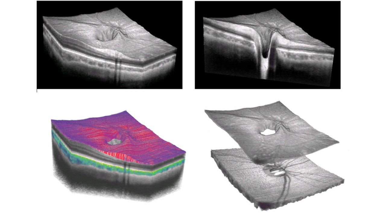
Retinal Layer Segmentation
Retinal OCTRetina is a layered tissue structure of neurons and synapses inside the eye that detect and process visual information. Optical coherence tomography (OCT) images the retina in-vivo with high-resolution, 3D visualization.
Retinal morphology is essentially related to function, as the tissue degradation in diseases such as glaucoma and age-related macular degeneration (AMD) leads to loss of vision that is often irrevocable.
Retinal Layer SegmentationSegmentation of retinal layers is a necessary step before quantitative shape analysis. Challenges to accurate segmentation include various imaging artefact and inconsistencies such as shadowing caused by intraretinal vasculature.
We use a robust graph-cut based algorithm that yields smooth and consistent layer boundary segmentation. The effect of imaging artefact and structural inconsistencies is mitigated by the three-dimensinoal nature of the algorithm in which the continuity of the segmented surface enforces correct delineation even for the edges with poor contrast.
 Publications
S. Lee, N. Fallah, F. Forooghian, A. Ko, K. Pakzad-Vaezi, A. B. Merkur, A. W. Kirker, D. A. Albiani, M. Young, M. V. Sarunic, M. F. Beg, “Comparative analysis of repeatability of manual and automated choroidal thickness measurements in non-neovascular age-related macular gegeneration,” Invest. Ophthalmol. Vis. Sci., 54(4), 2864-71 (2013)
S. Lee, M. F. Beg, M. V. Sarunic, “Segmentation of the macular choroid in OCT images acquired at 830 nm and 1060 nm,” European Conference in Biomedical Optics, Munich, Germany, 2013
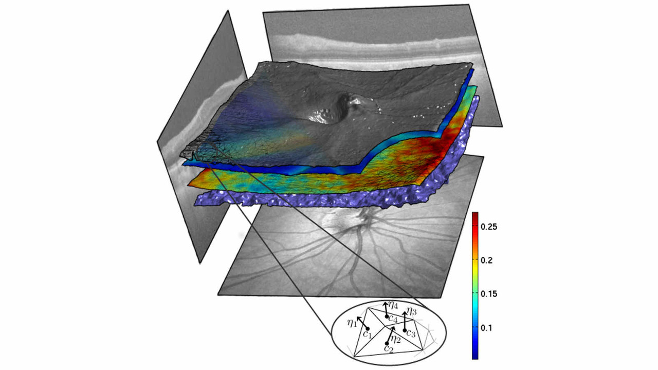
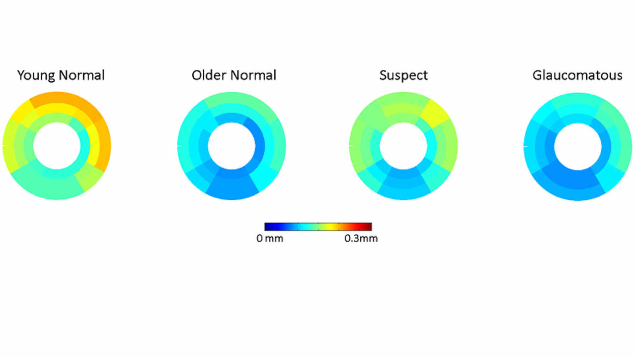
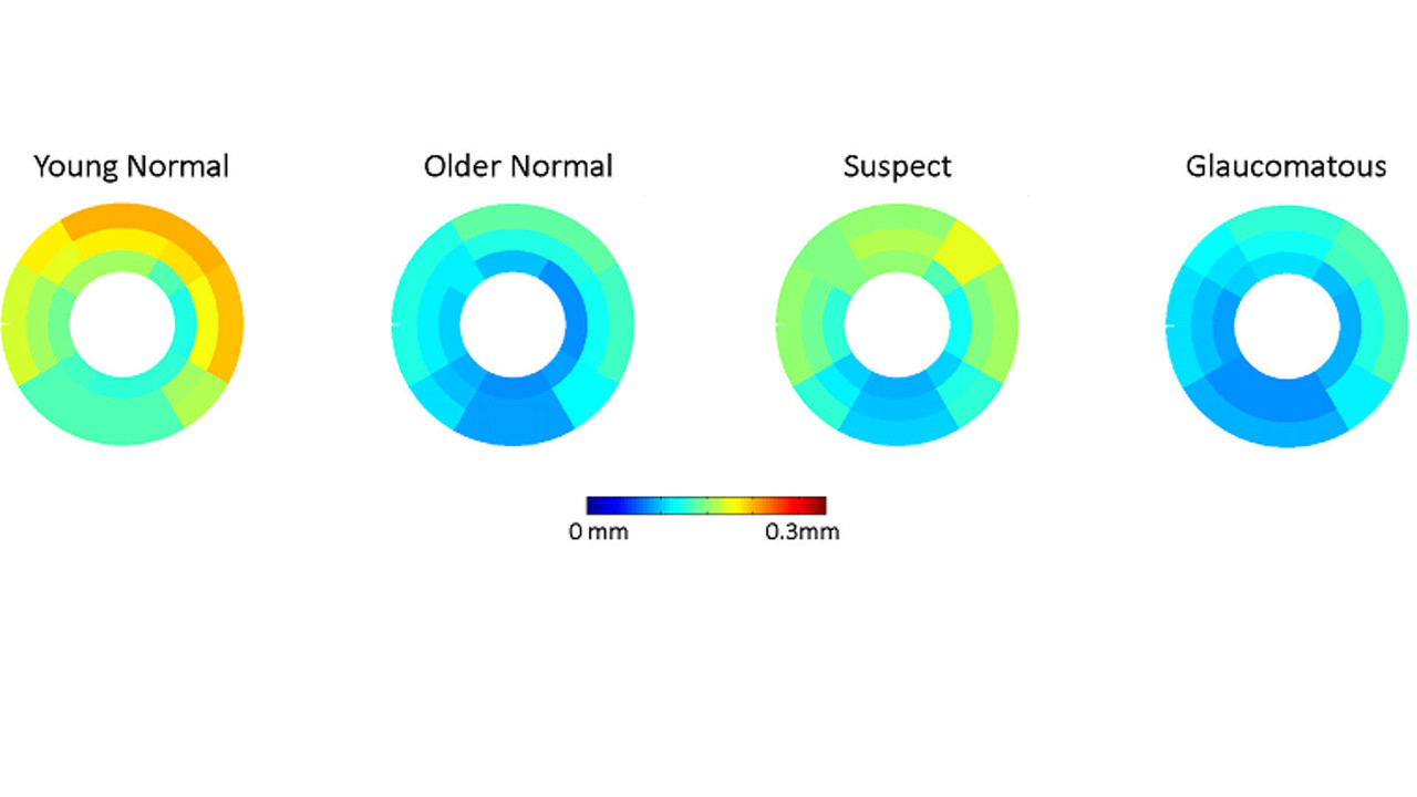
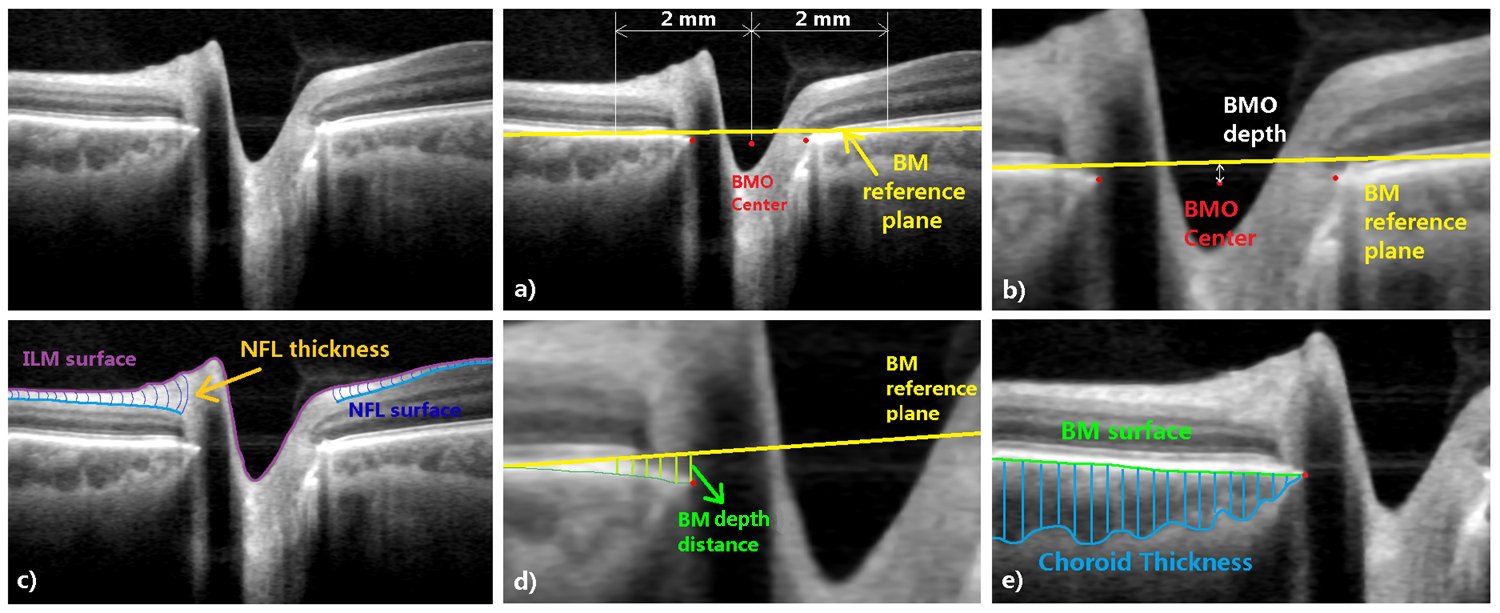
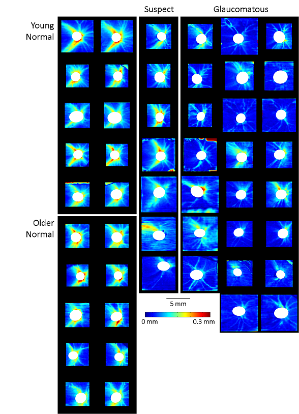
Optic Nerve Head Morphometrics
Glaucoma is the second leading cause of blindness in the world with irrevocable loss of vision due to damaged retinal cells. The main risk factor in glaucoma is increase intraocular pressure (IOP), and it has been suggested that structural degradation in the optic nerve head (ONH) region due to IOP is a critical event in the progression of the disease.
Optic Nerve HeadOptic nerve head (ONH) is the region in the posterior section of the eye where the retinal nerve fibers exit the globe toward the visual cortex of the brain. The axons are subject to various mechanical tensions and torsions, and a function of the ONH is to provide mechanical support for the axons in the presence of the IOP and cerebrospinal fluid pressure (CSF).
ONH MorphometricsOptical coherence tomography (OCT) enables in-vivo, 3D imaging of the ONH. The morphology of the ONH is an important indication of the structural change it goes through under various conditions, including glaucoma. In order to take full advantage of the rich information in 3D OCT, and perform comprehnsive quantitative shape analysis, an automated and robust morphometrics framework is required.
To understand the combined effect of glaucoma, myopia, and aging on the ONH, we imaged glaucoma patients and healthy controls with varying degrees of myopia and in different age groups. The acquired images were enhanced, segmented for key structures such as the retinal nerve fiber layer (RNFL), choroid, and Bruch's membrane opening (BMO), and measurements including the layer thickness, degree of surface bowing, and BMO dimension were made automatically. Mutliple regression was used to identify correlation between the shape parameters and the independent variables of age, degree of myopia, and severity of glaucoma.
 Publications
S. Lee, S. Han, M. Young, M. V. Sarunic, M. F. Beg, P. J. Mackenzie, "Optic nerve head peripapillary morphometrics in myopic glaucoma," Invest. Ophthalmol. Vis. Sci., 55(7):4378-93, 2014.
Retinal Surface Registration
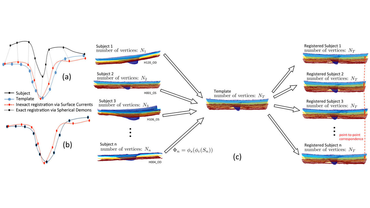
The challenge in quantifying the shape difference between two or more retinas from either a single subject (longitudinal) or multiple subjects (cross-sectional) is to establish anatomical correspondence between the retinas. The conventional approach is to define certain regions ("sectors") based on anatomical landmarks, scale, and orientation, but this often does not take into account individual variability among the subjects, and measurement values must be averaged over the regions with small, local changes becoming "averaged out" and not easily detectable.
Pointwise matching of retinal surfacesOur exact registration of a target retinal surface to a template retinal surface occurs in two stages: first, the surfaces are brought into close geometrical proximity by representing the surfaces as mathematical currents, and using the associated reproducing kernel Hilbert space norm to define and minimize the distance measure between the surfaces. This is followed by spherical demons registration which yields point-to-point correspondence between the surfaces.
PublicationsS. Lee, E. Lebed, M. Sarunic, M. F. Beg, "Exact Surface Registration of Retinal Surfaces from 3D Optical Coherence Tomography Images," IEEE Trans. Biomed. Eng., Accepted for publication (2014)
Image Registration and Segmentation
Large deformation diffeomorphic metric mapping (LDDMM) framework for computational anatomy.
Key paper:Beg, M. F., Miller, M. I., Trouvé, A., & Younes, L. (2005). Computing large deformation metric mappings via geodesic flows of diffeomorphisms. International journal of computer vision, 61(2), 139-157.
Image Acquisition
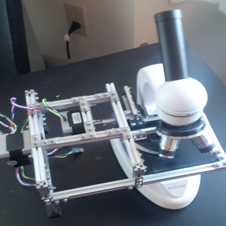
TB CamAn automated, low cost data acquisition system designed for deployment in impoverished nations, the TB Cam is intended to automate many aspects of diagnosing tuberculosis (TB) which are currently performed manually in many parts of the world. Designed to be a rugged, portable device, it is capable of performing automated 2D raster scans of a single microscope slide while also handling automatic focus control and safety control to prevent damaging slides while under analysis. Coupled with the ability to rapidly acquire dozens of images in seconds and then stitching them together for analysis and counting the number of bacteria detected, this device has the potential to become an extremely useful tool for reducing lead time on diagnosing potential TB patients. The final device also incorporates a touchscreen and simple UI for ease-of-use and direct control of the microscope slide positioning if manual control is desired.
Starting from humble beginnings as a prototype made from simple DSP/PLC chips and LEGO, the current design now uses aluminum t-slot extrusions and an AmScope microscope plus mechnical stage for the mechanical portion of the design, and a combination of a RaspBerry PI Model-B micro PC and custom hardware for motor control and analog to digital conversion (ADC). Work is currently underway to further drive down material costs through the use of 3D printing mechnical parts for the overall design, allowing for a complete turnkey solution for production and assembly of a complete, low cost device unimpeded by royalty fees or licensing constraints.
Quality Check Visualizations
Here we present the visualizations for the quality control of the processed data within our pipelines for the segmentation, registration and feature extraction tasks. Each link opens a new .pdf file presenting the corresponding processed data for all the subjects used in a particular study for which the publication is provided as well.
Publications:
