
Techniques
Electrophysiology
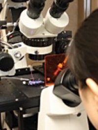
We perform different configurations of patch clamp (whole cell, inside-out etc.) or two-electrode voltage clamp using iPSC-CMs, Xenopus oocytes and mammalian cell lines. We also use sharp electrodes to record from isolated zebrafish hearts. We record both macroscopic and microscopic currents in a number of K+ channels using voltage clamp, and action potentials and voltage transients using current clamp. We also record in vivo ECG from adult and larval zebrafish.
CRISPR-Cas9 gene-editing
We are using CRISPR-Cas9 technology to engineer point mutations in the hERG channel gene in iPSC-derived cardiomyocytes (hiPSC-CMs) and in zebrafish hearts. Our approach utilizes the homology directed repair or non-homologous end joining mechanisms to incorporate a nucleotide changes following a targeted Cas9-induced double stranded break. Functional characterization involves electrophysiology, optical mapping and ECG.

HIPSC-CM ACTION POTENTIALS
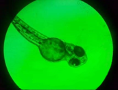
ZEBRAFISH CRISPR EDIT SCREEN

ZEBRAFISH ECG
Optical mapping

We collaborate with the Tibbits lab in the Molecular Cardiac Physiology Group to study iPCS-derived cardiomyocytes or ex vivo zebrafish whole hearts using optical mapping. Dual camera mapping enables simultaneous visualization of calcium transient and membrane voltage dynamics to report on cardiac electrophysiological function.
Voltage Clamp Fluorimetry (VCF)
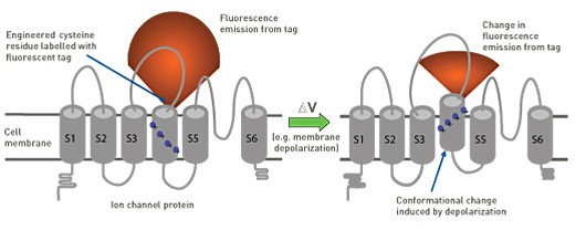
- Conformational changes alter the fluorescent tag’s environment and this changes its fluorescence emission
- This enables direct observation of the molecular mechanisms of channel activity in real time
- Simultaneous measurement of fluorescence emission changes along with ionic current provides an encompassing and unparalleled report of channel gating processes
- The fluorescence approach enables visualization of conformational changes even in closed or inactive ion channels, which are “invisible” when measuring ionic current alone
Cut-Open Oocyte Voltage Clamp (COVC)
COVC is a modified Xenopus oocyte voltage clamp configuration that allows for very rapid voltage control of the membrane. This enables us to measure rapid events, such as gating currents produced by voltage sensor domain movement. Combined with simultaneous fluorescence measurements, this set-up provides a powerful system with which to investigate ion channel behaviour.
Lanthanide-Based Resonance Energy Transfer (LRET)
LRET enables quantitative measurement of distances between two sites within the channel and how these change with protein dynamics. One fluorescent probe is attached within the channel as a donor and a second probe is engineered as an acceptor. The transfer of energy from donor to acceptor is measured upon excitation of the donor. The transfer of energy is directly related to the distance between the donor and acceptor. This provides a quantitative evaluation of the dynamics of channel function. We are using LRET to measure dyanamic distance changes in hERG channels.
Protein Biochemistry
We have wide ranging experience performing SDS-PAGE and western blot analysis of wild-type and mutant ion channels expressed in different cell types. These data are useful in understanding how proteins are trafficked to the cell membrane.
Site-Directed Mutagenesis
In order to investigate the molecular mechanisms underlying ion channel function, we engineer single point mutations, or deletions/insertions of multiple amino acids, into the protein. Comparisons amongst different ion channels enables the identification of important sites that may govern a particular phenotype in a given channel. PCR-based site-directed mutagenesis provides a highly efficient method to generate mutant channel constructs for these investigations.