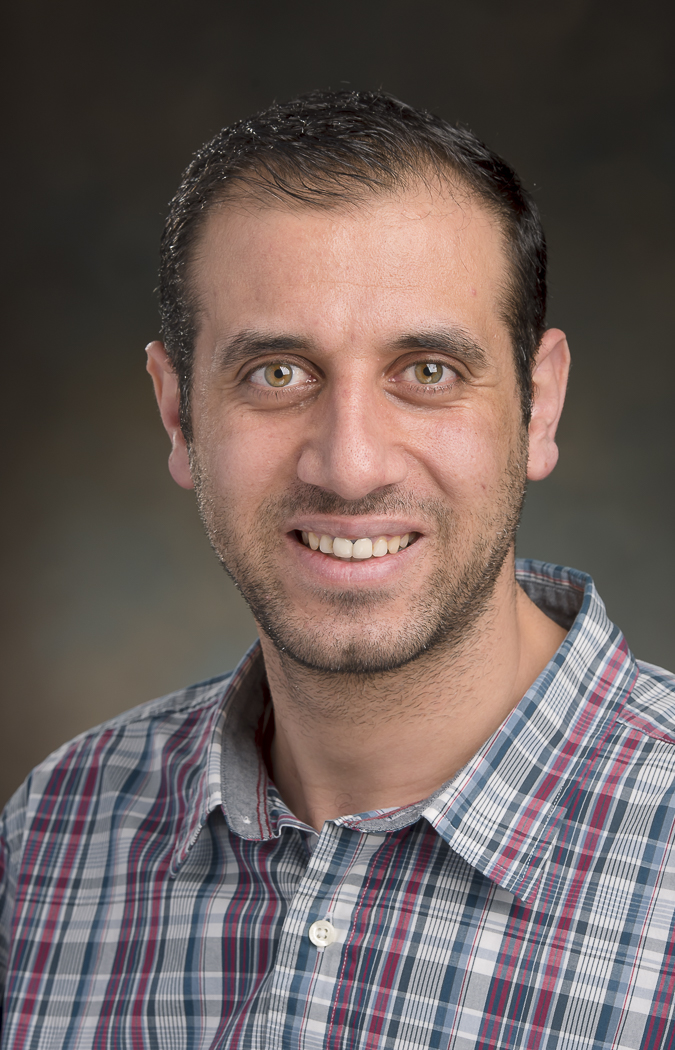
Ismail M. Khater
Research Summary
Ismail M. Khater, Simon Fraser University
Computational Complex Network Analysis for Single Molecule Localization Super-resolution Microscopy:
My primary research interest lies at the intersection of biomedical image analysis and computing science. In particular, my work has primarily focused on utilizing complex network models and graph theory measures to analyze big-data that comes from super-resolution microscopic images. I have a particular interest in studying prostate cancer and cardiac diseases. This interest is motivated by the recent development of super-resolution microscopy which allows the researcher to go beyond the diffraction limit of the light microscopy. The current methods and tools are typically computationally inefficient which limits the region of interest to small 2D areas and are limited to crude global measures. Our research, on the other hand, encompasses both 1) Building tools to study/examine particular clinical questions regarding diseases as well as design and implement algorithms to test clinical hypotheses. 2) Developing new and robust tools using parallel programming, GPUs, and high performance computing (e.g. cluster computing).
In 2014, Betzig et al. have won the Nobel prize in chemistry [1]. They were honored for bringing “optical microscopy into the nanodimension” or what is called super-resolution microscopy. Super-resolution allow images to be taken at higher resolution than the diffraction limit of the conventional light microscopy. This unprecedented imaging advancement is enabling us to study subcellular compartments at the molecular level. Defining the nano- and meso-scale architecture that exists below the 200 nm diffraction limit of visible light is key to understanding how the cell works and has driven some recent amazing progress in super-resolution microscopy.
Our research focuses on studying 3D protein localization from super-resolution images to improve our understanding of prostate cancer. We are developing novel quantitative approaches to model and analyze the large datasets of 3D point localizations. We model millions of X,Y,Z “blink” locations (i.e. 3D point clouds) as networks with virtual connections constructed between the points. This network representation is well-established for modelling brain, social, and computer networks [7,8]. Our group is the first to leverage network analysis in 3D of prostate cancer super-resolution microscopic images [2-6].
Our computational approach is based on studying the protein-to-protein interaction at various proximity scales to extract bio-signatures that leads to segmentation, classification, matching, and identification of the different categories of the subcellular structures from different prostate cancer populations. Existing methods to analyze super-resolution data, e.g. [9-11], do not consider the whole cell, are limited to a 2D small region of interest (ROI), and only provide global measures at the ROI level. Our approach, on the other hand, is the first to deal with the whole super-resolution cell of 3D point cloud and obtain multi-scale measures for every point/protein (i.e. per-point features) in the data. We are already witnessing new exciting insights [2] into prostate cancer subcellular protein configurations that improve our understanding of the disease and drug design.
REFERENCES
[1] The-Scientist.com, October 8, 2014. http://www.the-scientist.com/?articles.view/articleNo/41169/title/Nanoscopy-Wins-Nobel/
[2] Khater IM, Meng F, Nabi IR, Hamarneh G. Super resolution network analysis defines the molecular architecture of caveolae and caveolin-1 scaffolds. Scientific Reports, 8(1):9009, 2018
[3] Khater IM, Scriven D, Moore E, Hamarneh G. Sub-cellular network analysis of ryanodine receptor positioning in control and phosphorylated states. In IEEE Computing in Cardiology (IEEE CinC), pages 821–824, 2016.
[4] Khater IM, Meng F, Nabi IR, Hamarneh G. Identifying Sub-Cellular Domains in PC3 cells by Analysing Caveolin-1 Proteins from Nanoscopy Super-Resolution Data, NanoLytica 2015, Posters, Burnaby, Canada
[5] Meng F, Khater IM, Hamarneh G, and Nabi IR. Super-resolution analysis of Caveolae and Cav1 scaffolds in Prostate Cancer Cells, Canadian Cytometry and Microscopy Association (CCMA) 2015, Toronto, Canada
[6] Nabi IR, Meng F, Khater IK, Hamarneh G. Super-resolution analysis of Caveolae and Cav1 scaffolds in Prostate Cancer Cells, Advanced Structural & Chemical Imaging (ASCI) Symposium 2015, Pullman, Washington
[7] Brown CJ, Miller SP, Booth BG, Andrews S, Chau V, Poskitt KJ, Hamarneh G. Structural network analysis of brain development in young preterm neonates. NeuroImage 2014;101:667-80.
[8] Newman M. Networks: An Introduction: Oxford University Press; 2010.
[9] Owen D, Rentero C, Rossy J, Magenau A, Williamson D, Rodriguez M et al. PALM imaging and cluster analysis of protein heterogeneity at the cell surface. Journal of biophotonics. 2010 Jul;3(7):446-454.
[10] Andronov L, Lutz Y. Vonesch J.-L., Klaholz B. P. SharpViSu: integrated analysis and segmentation of super-resolution microscopy data. Bioinformatics. 2016
[11] Levet F, Hosy E, Kechkar A, Butler C, Beghin A, Choquet D, Sibarita J. SR-Tesseler: a method to segment and quantify localization based super-resolution microscopy data. Nat Methods 2015; 12, 1065–1071
