sfu.ca
F
T
I
CS Intranet
MENU
Faculty of Applied Sciences
School of Computing Science
sign in
SFU Mail
go
SFU
Canvas
Search
This Site
SFU.ca
About
Message From the Director
Industry Relations
Job Opportunities
Diversity in CS
Contact Us
CS Intranet
People
Leadership
Faculty
Staff
Future Students
Undergraduates
Graduates
Current Students
Undergraduates
Graduates
Student Life
Research
Areas
Labs
Centres & Institutes
Chairs
Awards
Seminars
News & Events
News
Events
Support
CSIL Linux/UNIX FAQ
CSIL
Grads & Researchers
Guides & Tips
School of Computing Science
News & Events
School of Computing Science
About
Message From the Director
Industry Relations
MPCS Advisory Council
Job Opportunities
Diversity in CS
Diversity Committee Webinars
Awards and Project Presentations
2025 Explore Computing Science Research Workshop
Contact Us
CS Intranet
People
Leadership
Faculty
Staff
Future Students
Undergraduates
Graduates
Current Students
Undergraduates
Program Requirements
Forms
Policies
Co-op
Student Resources
Undergraduate Student Research Award (USRA)
Academic Enhancement Program (AEP)
AEP in the News!
Computing Science Peer Tutoring
Meet your CS Peer Tutors
Research Opportunities
Undergraduate Project Showcase
Frequently Asked Questions
Graduates
Program Requirements
Policies
Forms
Financial Support
Resources
Supervisory Committee
Thesis & Defence
Internships & Co-op
MPCS Co-op
Industrial Internships
Teaching Assistantships
Student Life
Research
Areas
Labs
Trustworthy Artificial Intelligence (TAI) Lab
Centres & Institutes
Chairs
Awards
Nominations for CS Excellence in Teaching Awards
Best Paper Award
Seminars
Distinguished Lecture Series
Industry Talks Series
VCR/AI
News & Events
News
Events
ICPC Programming Contest
About ICPC
ICPC PACNW | SFU
Support
CSIL Linux/UNIX FAQ
CSIL Linux/Unix Known Issues
Network Lab FAQ
CSIL
Remote Access
CSIL Windows systems
Known Issues
CSIL Linux/UNIX FAQ
Basic Linux Command Line Survival
Known Issues
How to kill Unix Processes
Grads & Researchers
Guides & Tips
Print, Photocopy & Scan
News & Events
Latest News
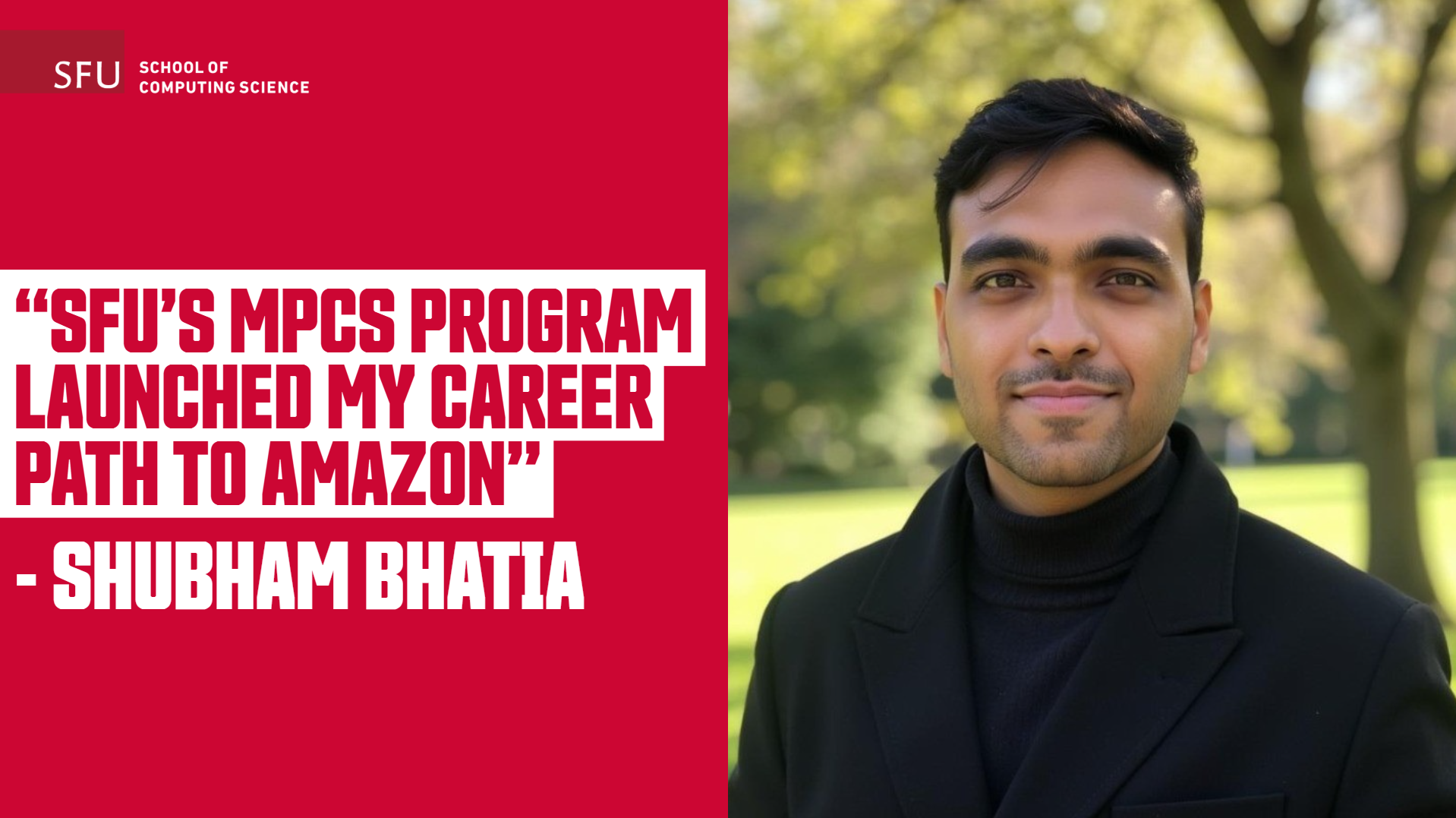
Shaping the Future with Big Data: Shubham Bhatia’s MPCS Experience at SFU
Read More
New AI Tool Lean Finder Helps Mathematicians Discover Theorems Faster and Smarter
Read More
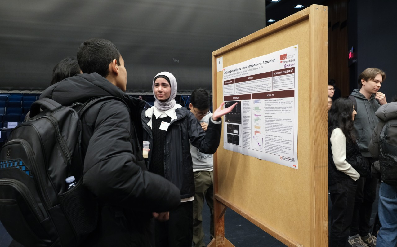
Celebrating Innovation and Discovery at CS Research Day 2025
Read More
Read more news
Upcoming Events
See more events
About
Overview
Message From the Director
Industry Relations
Overview
MPCS Advisory Council
Job Opportunities
Diversity in CS
Overview
Diversity Committee Webinars
Awards and Project Presentations
2025 Explore Computing Science Research Workshop
Contact Us
CS Intranet
People
Overview
Leadership
Faculty
Staff
Future Students
Overview
Undergraduates
Graduates
Current Students
Overview
Undergraduates
Overview
Program Requirements
Forms
Policies
Co-op
Student Resources
Undergraduate Student Research Award (USRA)
Academic Enhancement Program (AEP)
AEP in the News!
Computing Science Peer Tutoring
Meet your CS Peer Tutors
Research Opportunities
Undergraduate Project Showcase
Frequently Asked Questions
Graduates
Overview
Program Requirements
Policies
Forms
Financial Support
Resources
Supervisory Committee
Thesis & Defence
Internships & Co-op
MPCS Co-op
Industrial Internships
Teaching Assistantships
Student Life
Research
Overview
Areas
Labs
Overview
Trustworthy Artificial Intelligence (TAI) Lab
Centres & Institutes
Chairs
Awards
Overview
Nominations for CS Excellence in Teaching Awards
Best Paper Award
Seminars
Overview
Distinguished Lecture Series
Industry Talks Series
VCR/AI
News & Events
Overview
News
Events
Overview
ICPC Programming Contest
About ICPC
ICPC PACNW | SFU
Support
Overview
CSIL Linux/UNIX FAQ
Overview
CSIL Linux/Unix Known Issues
Network Lab FAQ
CSIL
Overview
Remote Access
CSIL Windows systems
Known Issues
CSIL Linux/UNIX FAQ
Basic Linux Command Line Survival
Known Issues
How to kill Unix Processes
Grads & Researchers
Guides & Tips
Overview
Print, Photocopy & Scan
SFU.CA
Search
Search
This Site
SFU.ca
SFU Mail
go
SFU
Canvas
close