|
Skin Lesion Analysis (Pigment
Network Detection)
Problem:
Motivation:
Abstract:
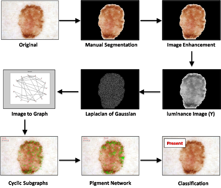
We describe a novel approach to detect and visualize
pigment network structures in dermoscopic images, based on
the fact that the edges of pigment network structures form
cyclic graphs which can be automatically detected and
analyzed. First we perform a preprocessing step of image
enhancement and edge detection. The resulting binary edge
image is converted to a graph and the defined feature
patterns are extracted by finding cyclic subgraphs
corresponding to skin texture structures. We filtered these
cyclic subgraphs to remove other round structures such as
globules, dots, and oil bubbles, based on their size and
color. Another high-level graph is created from each
correctly extracted subgraph, with a node corresponding to a
hole in the pigment network. Nodes are connected by edges
according to their distances. Finally the image is
classified according to the density ratio of the graph. Our
results over a set of 500 images from a well known atlas of
dermoscopy show an accuracy of 94.3% on classification of
the images as pigment network Present or Absent.
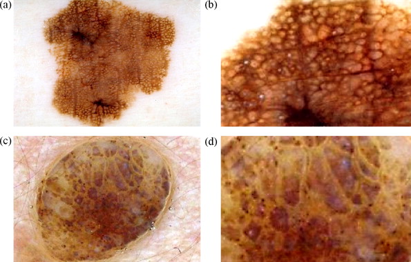
Fig. 1. (a) Present: a
lesion containing a pigment network. (b) Enlarged pigment
network. (c) Absent: an image of a lesion without
pigment network. (d) Enlarged Absent image.
Method
Overview:
Detecting pigmented network
is a crucial step for melanoma diagnosis. In this paper, we
present a novel graph-based pigment network detection method
that can find and visualize round structures belonging to
the pigment network.
more details...
Results:
Step by step results:
More Results:
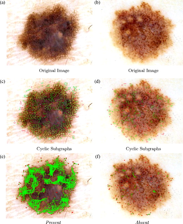
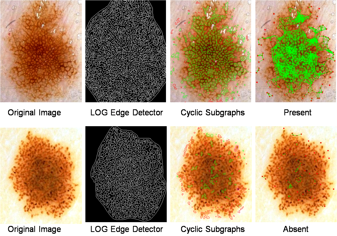
Results of applying our approach to two
Present and Absent dermoscopic images: (a and b)
are skin lesions, (c and d) show cyclic subgraphs, the green
meshes represent potential holes of the pigment network and
red meshes could not pass the test of belonging to the
pigment network, and (e and f) visualize the pigment network
over the image.
Conference Publication (PDF,
Presentation PPT):
M. Sadeghi, M. Razmara, M. Ester, T. K. Lee, M. S. Atkins,
ōGraph-based Pigment Network Detection in Skin Imagesö, SPIE --
Medical Imaging 2010, Vol 7623, Feb. 2010
Journal Publication (PDF): Maryam
Sadeghi, Majid Razmara, Tim K. Lee and M. Stella Atkins, ōA novel method
for detection of pigment network in dermoscopic images using
graphsö, Special Issue: Skin Cancer Imaging Computerized
Medical Imaging and Graphics Journal, in press, 2010,
doi:10.1016/j.compmedimag.2010.07.002
|