x
close

Magnet Safety
Simon Fraser University has a comprehensive Radiation Safety Program. The use of magnetic instrumentation falls under the Radiation Safety Program, whose primary objective is to facilitate and ensure the safe use of magnetic devices in research, teaching, and the environment.
For more information, contact the Program Manager, Radiation Safety via email: rso-info@sfu.ca, or ph: 778-782-3633
With Nuclear Magnetic Resonance (NMR), MRI and Superconducting magnetic equipment in general there are a number of unique safety concerns. Consultation with EHS is required to determine specific hazards for each facility housing such magnetic sources in order to identify hazardous areas and review safety measures. Magnet instrumentation registration has been added to the SFU laboratory hazard inventory system as Non-Ionizing Radiation (NIR) equipment.
Magnetic Fields
Non-ionizing Radiation (NIR) refers to any type of electromagnetic radiation that does not carry enough energy to ionize atoms or molecules. NIR is part of the electromagnetic (EM) spectrum and is characterized by waves. NIR differs from ionizing radiation by having lower quantum energies, and having different biological effects. NIR shares the wave characteristics of ionizing radiation and can be described by wavelength, frequency and energy. When compared to ionizing radiation, NIR consists of longer, less frequent and less energetic properties, but can still inflict a good deal of damage.
The intensity of a magnetic field is usually measured in Tesla or millitesla (T or mT) or Gauss (G).
1 T = 10,000 G 1 mT = 10 G 0.5 mT = 5 G
Note: Household magnets have strength on the order of several tens of millitesla (mT)
Static Magnetic Fields
- A static magnetic field is a force field created by a magnet or charges that move in a steady flow to produce direct current (DC). They exert an attractive force on metallic objects containing iron, nickel, or cobalt, for example.
- Sources of static magnetic fields found at SFU include Nuclear Magnetic Resonance (NMR) equipment, spectroscopy systems, ion pumps, MRI, bend magnets, superconducting magnets and cryostats.
- Important: Static fields can erase data stored on magnetic media or on the strips of banking cards.
- Magnets which generate a static magnetic field have been added to the lab hazardous inventory as Non-Ionizing Radiation (NIR) equipment.
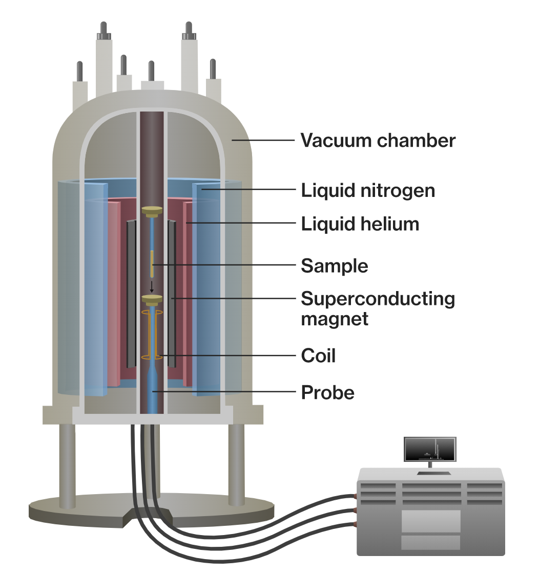
Superconducting Magnets
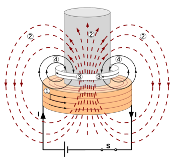
1. Coil current
2. Upward fast increasing magnetic field
3. Induced current
4. Magnetic field of the induced current
Superconducting Magnets consist of electromagnets made from coils of supercondcuting wire. They must be cooled to cryogenic temperatures during operation. The wire can conduct very high electric currents when in its superconducting state, much higher than ordinary wires, generating an intense magnetic field.
Superconducting magnets are used in NMR spectrometers, mass spectrometers, particle accelerators and MRI scanners.
Superconducting magnets pose unique safety concerns including cryogen safety, strong magnetic fields, and the potential to create an oxygen deficient atmosphere. The most serious hazards exists during the magnet start-up. Once the magnetic fields have been established and are operational the hazards are low as long as operators and personnel understand the proximity limits and procedures to follow when approaching the magnet.
Nuclear Magnetic Resonance
The NMR system uses a static magnetic field to align nuclear spins in the magnetic field to maximize the NMR signal. NMR spectroscopy exploits the magnetic properties of atomic nuclei to obtain information about the structure, dynamics and chemical environment of molecules.
NMR systems consist of superconducting magnets which produce core fields from 0.15 T to 20 T. These fields decrease in intensity with the increase of the core distance. NMR systems in research are more powerful than medical devices, but their fields are characterized by having a smaller volume, being more focused, and falling off quickly, making personnel protection an easy provision.
Magnetic Resonance Imaging

In Radiology the MRI technique is used to generate images of organs in the body for diagnostic purposes. MRI scanners employ large, powerful magnets which produce strong magnetic fields. Patients are placed inside an MRI machine for medical procedures. The magnetic field temporarily forces protons in the body to align with the field. When a radiofrequency current is pulsed through a patient, the protons are stimulated, and spin out of equilibrium, straining against the pull of the magnetic field. When the radiofrequency field is turned off, the MRI sensors are able to detect the energy released as the protons realign with the magnetic field. Faint signals are produced in this process and are used to create cross-sectional MRI images. The radio waves are used to send signals to the body and receive signal from the body in return. The signals are converted into images by computer software.
Clinical magnets are superconducting magnets and require liquid helium cooling. The MRI magnetic field strength ranges from 0.15 T to 4 T. Superconducting magnets at 1.5 T and above allow functional brain imaging and MR spectroscopy with improved time and spatial resolution. Such magnets have additional challenges from radiofrequency (RF) heating of the subject.
Time-varying Magnetic Fields
- Time-varying magnetic fields are magnetic fields that reverse their direction at a regular frequency. They can induce an electric current in a conductor present in this field as well as in a human body.
- Time-varying magnetic fields are produced by devices using alternating current (AC) such as cellular telephone antennas, microwaves etc.
- Induced currents in the body can cause local heating and possible burns.
F T I